LSU Tiger Stadium 360º View

[vrview img=”https://kermitmurray.com/images/stadium360.JPG” ]
Murray Mass Spectrometry Group
Research group of Kermit Murray at Louisiana State University where we use lasers for sampling and imaging and study the chemistry and physics of laser ablation.
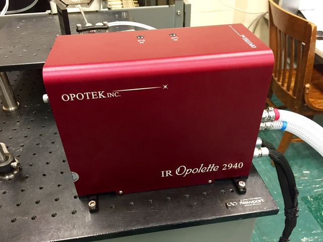
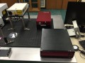
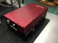
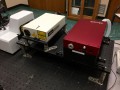
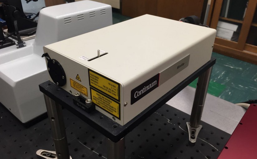
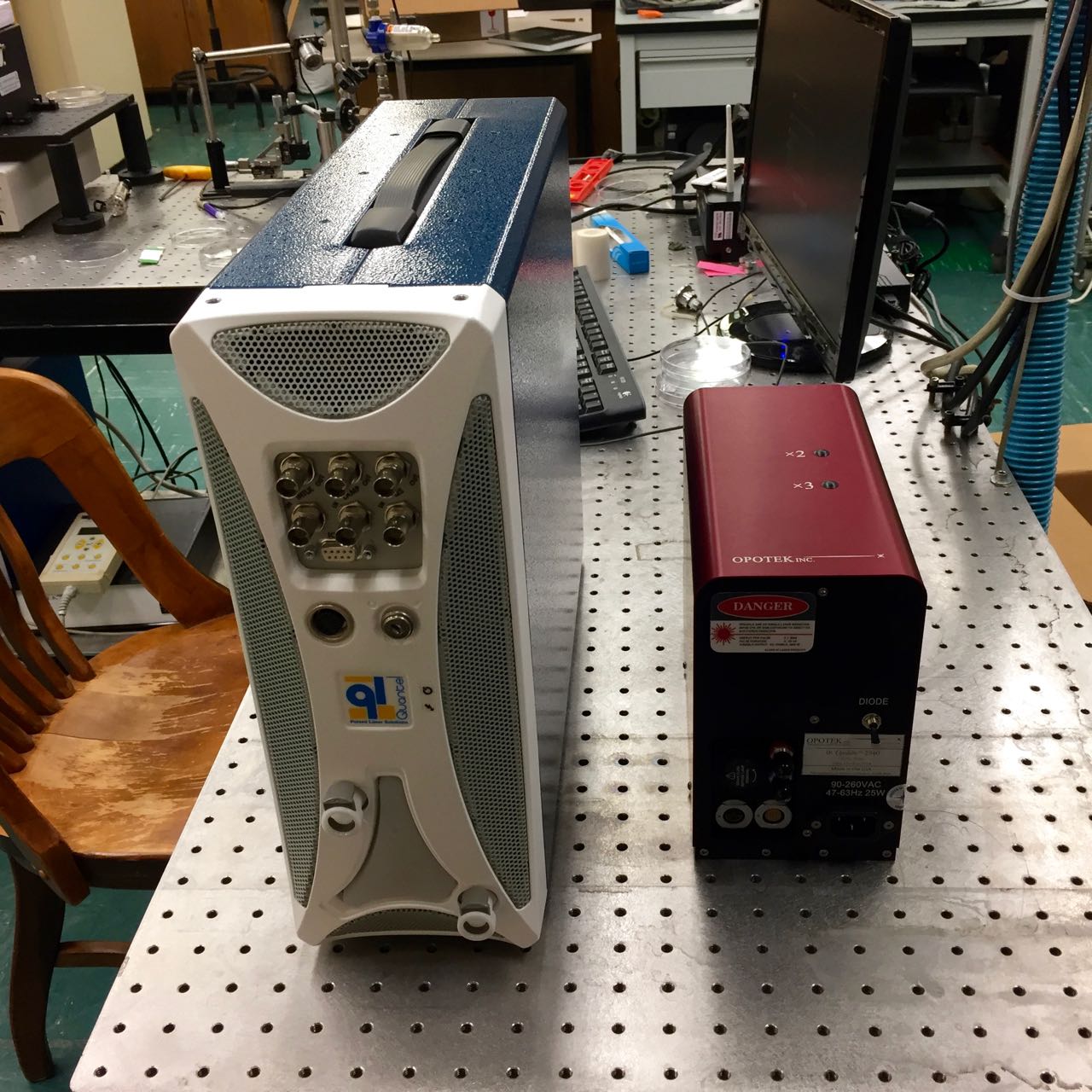
Another laser and the second from OPOTEK this year: an IR Opolette 2940. It is fixed wavelength at 2940 nm for no other reason than that is the Er:YAG laser wavelength. It is possible to manually adjust the internal optics to generate light from 2700 to 3100 nm.
This laser has its roots in the STTR grant that I had with OPOTEK starting in 2001 (awarded when I was at Emory but moved immediately to LSU in the first year of Phase I). Our goal was to build an OPO with the capabilities of the Mirage 3000B but in a smaller package.
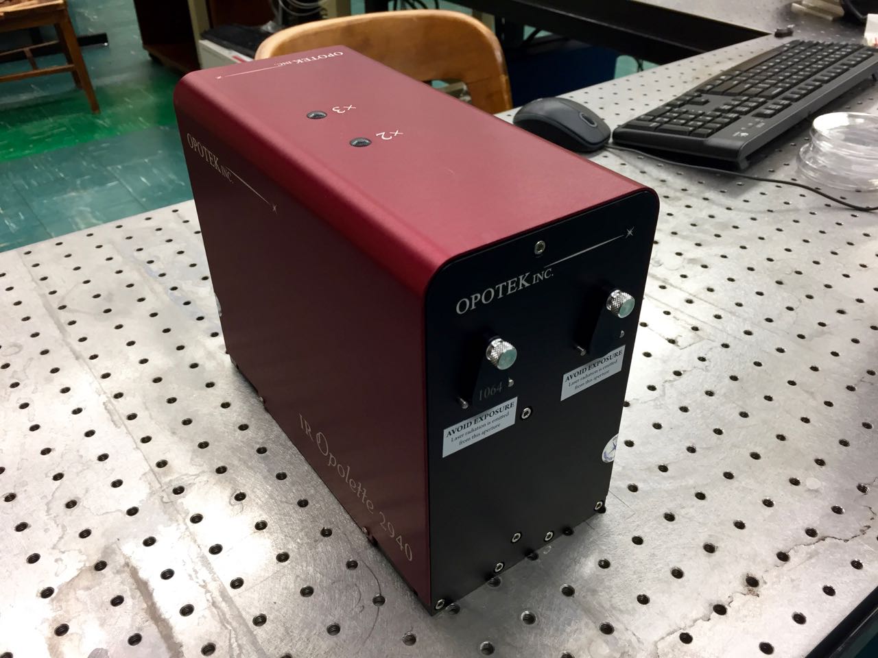
The left port gives access to the Nd:YAG laser fundamental at 1064 nm and the right port is the 2940 nm mid-IR at about 3 mJ per pulse at 20 Hz. Plenty of energy when focused to efficiently ablate thin films (similar to our wavelength tunable IR Opolette which you can see ablating things here.)
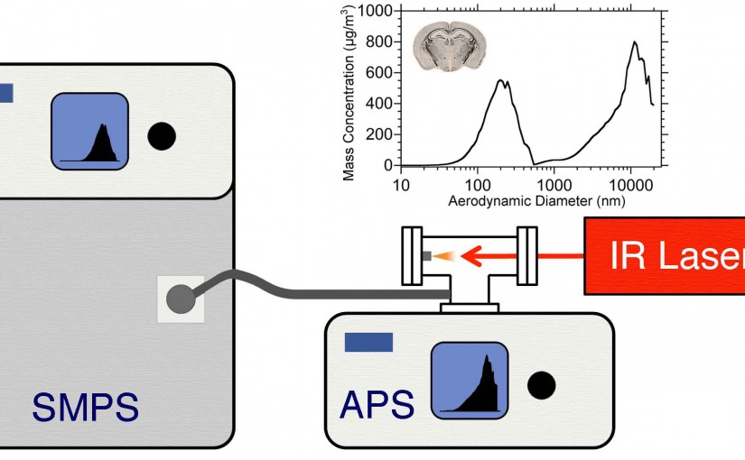
F. Cao, F. Donnarumma, K.K. Murray, “Particle size measurement from infrared laser ablation of tissue,” Analyst. 141 (2016) 183–190. doi:10.1039/C5AN01765C.
Abstract: The concentration and size distribution were measured for particles ablated from tissue sections using an infrared optical parametric oscillator laser system. A scanning mobility particle sizer and light scattering particle sizer were used in parallel to realize a particle sizing range from 10 nm to 20 μm. Tissue sections from rat brain and lung ranging in thickness between 10 and 50 μm were mounted on microscope slides and irradiated with nanosecond laser pulses at 3 μm wavelength and fluences between 7 and 21 kJ m−2 in reflection geometry. The particle size distributions were characterized by a bimodal distribution with a large number of particles 100 nm in diameter and below and a large mass contribution from particles greater than 1 μm in diameter. The large particle contribution dominated the ablated particle mass at high laser fluence. The tissue type, thickness, and water content did not have a significant effect on the particle size distributions. The implications of these results for laser ablation sampling and mass spectrometry imaging under ambient conditions are discussed.


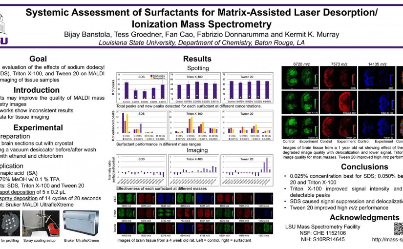
B. Banstola, T. Groedner, F. Cao, F. Donnarumma and K. K. Murray, Louisiana State University, Department of Chemistry, Baton Rouge, LA
K. K. Murray, , C. A. Seneviratne, and S. Ghorai, “High Resolution Laser Mass Spectrometry Bioimaging” Methods 104 (2016) 118–126; doi:10.1016/j.ymeth.2016.03.002
Mass spectrometry imaging (MSI) was introduced more than five decades ago with secondary ion mass spectrometry (SIMS) and a decade later with laser desorption/ionization (LDI) mass spectrometry (MS). Large biomolecule imaging by matrix-assisted laser desorption/ionization (MALDI) was developed in the 1990s and ambient laser MS a decade ago. Although SIMS has been capable of imaging with a moderate mass range at sub-micrometer lateral resolution from its inception, laser MS requires additional effort to achieve a lateral resolution of 10 lm or below which is required to image at the size scale of single mammalian cells. This review covers untargeted large biomolecule MSI using lasers for desorption/ionization or laser desorption and post-ionization. These methods include laser microprobe (LDI) MSI, MALDI MSI, laser ambient and atmospheric pressure MSI, and near-field laser ablation MS. Novel approaches to improving lateral resolution are discussed, including oversampling, beam shaping, transmission geometry, reflective and through-hole objectives, microscope mode, and near-field optics.
https://kermitmurray.com/documents/murray_2016.pdf