ASMS 2026: Laser Bioanalytics Corporate Poster
Murray Mass Spectrometry Group
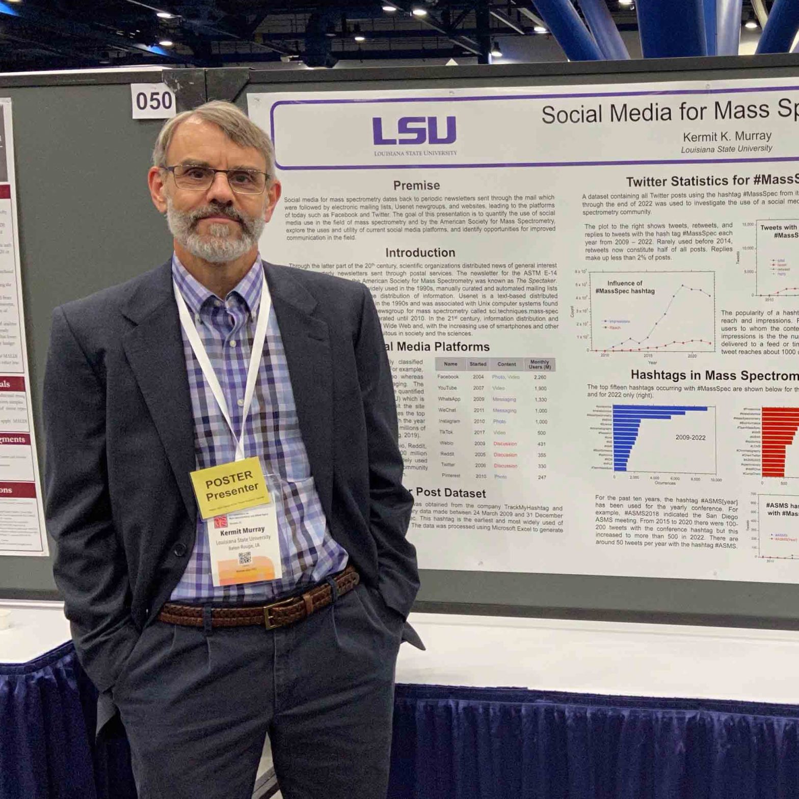
Research group of Kermit Murray at Louisiana State University where we use lasers for sampling and imaging and study the chemistry and physics of laser ablation.
After 25 years in Baton Rouge, Professor of Chemistry Kermit Murray has retired from LSU to work full time at his startup company Laser Bioanalytics LLC. The company is currently located in LSU Innovation Park, but will be moving to the Denver metro area in the summer of 2026.
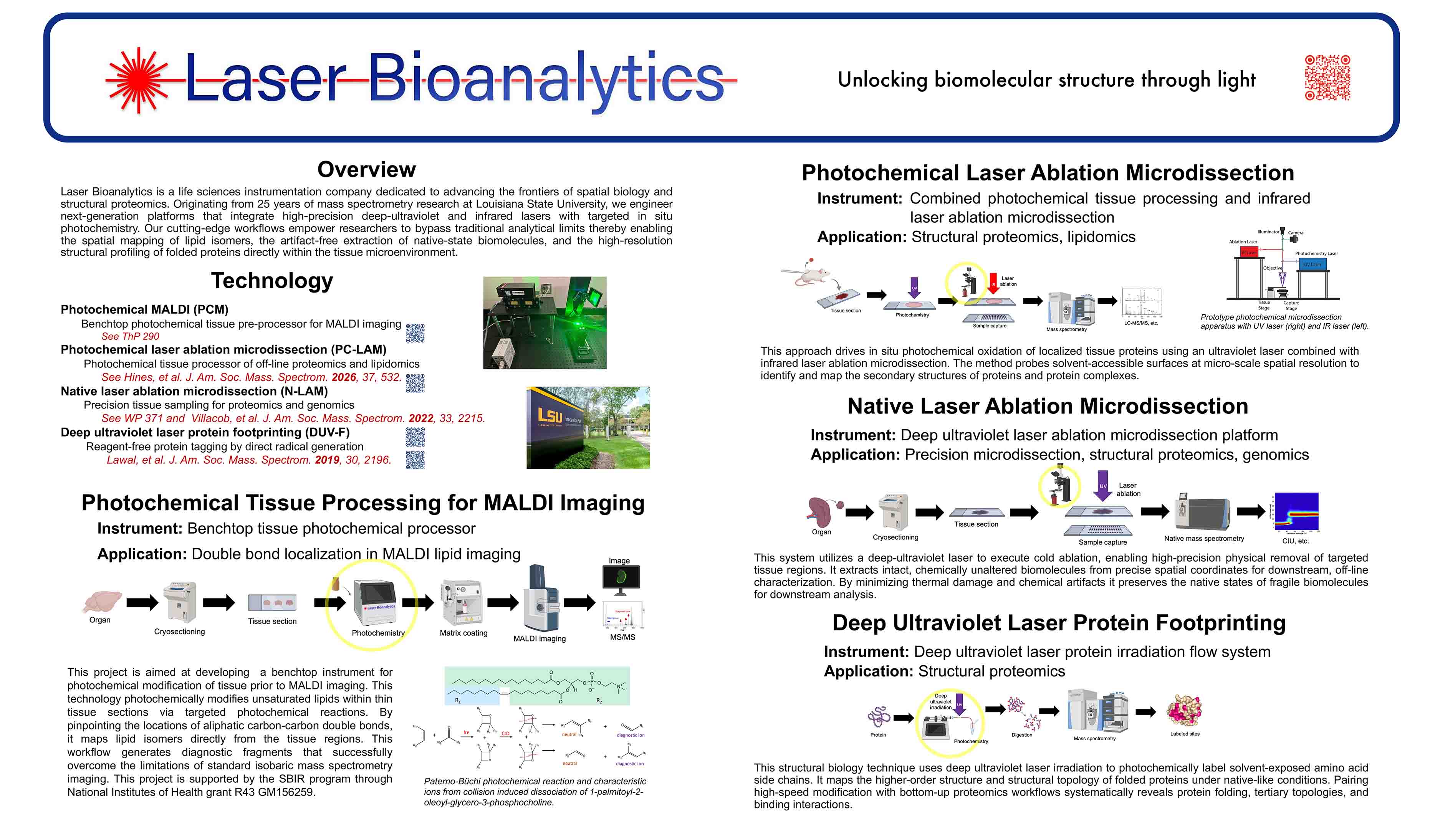
Laser Bioanalytics is a life sciences instrumentation company dedicated to advancing the frontiers of spatial biology and structural proteomics. Originating from 25 years of mass spectrometry research at Louisiana State University, the company engineers next-generation platforms that integrate high-precision deep-ultraviolet and infrared lasers with targeted in situ photochemistry. The cutting-edge workflows empower researchers to bypass traditional analytical limits thereby enabling the spatial mapping of lipid isomers, the artifact-free extraction of native-state biomolecules, and the high-resolution structural profiling of folded proteins directly within the tissue microenvironment.
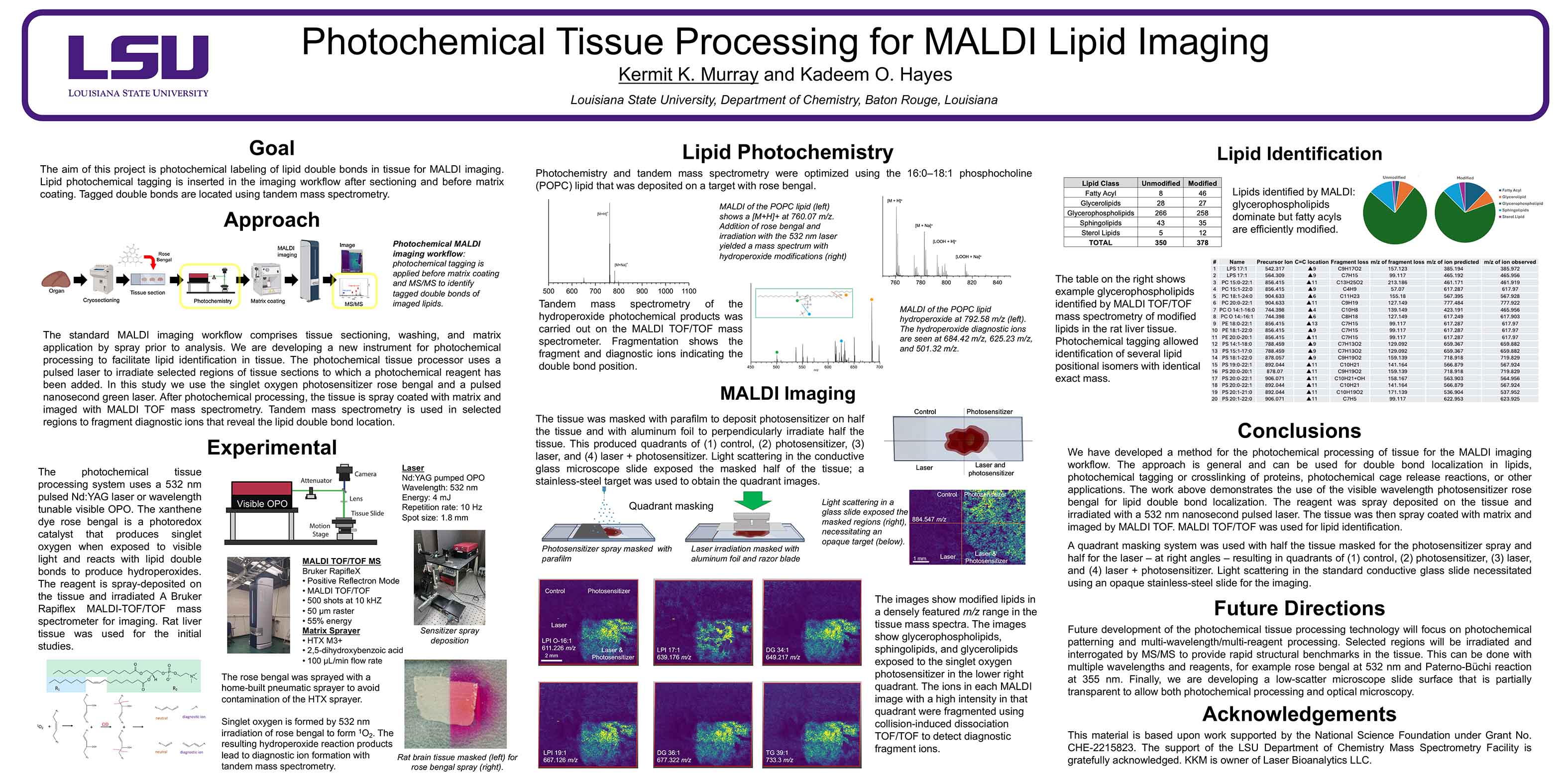
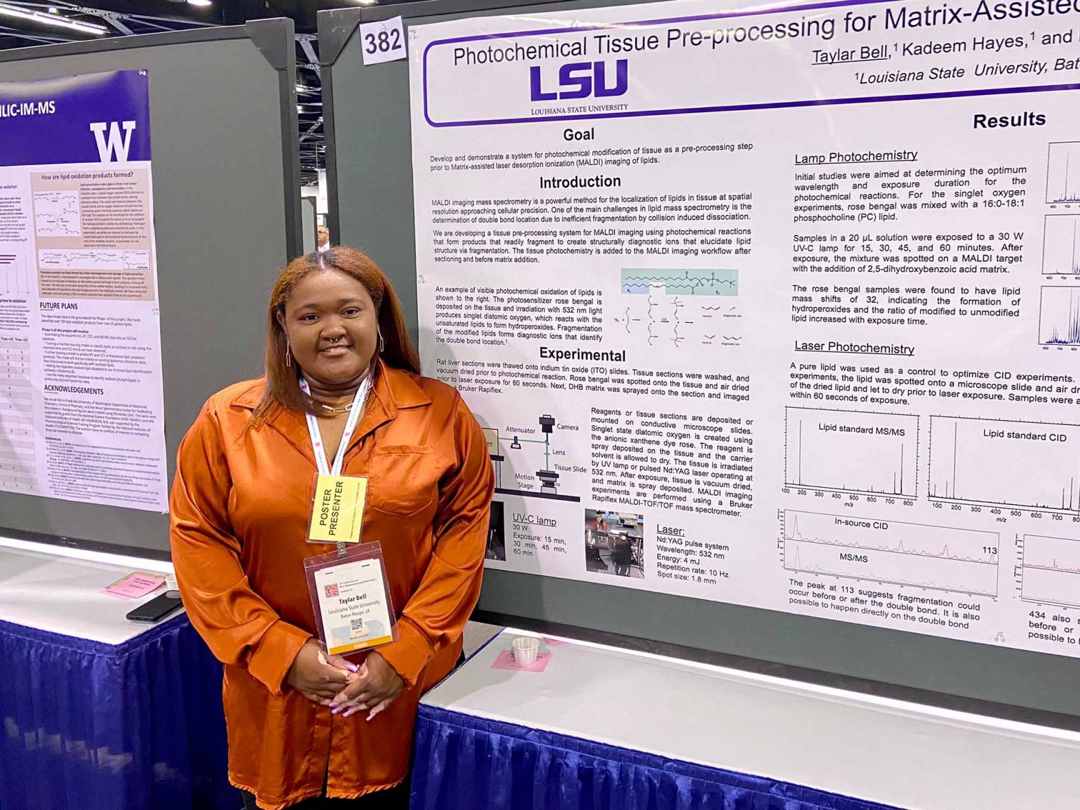
A photochemical tissue region modification system for the MALDI imaging workflow.
The standard MALDI imaging workflow comprises tissue sectioning, washing, and matrix application by spray or sublimation prior to analysis. We are developing a new instrument for tissue photochemical processing that produces biomarker compounds to facilitate compound identification. The current study is aimed at developing a method for lipid double bond localization using MALDI imaging and tandem time-of-flight mass spectrometry. The photochemical tissue processor uses a pulsed laser to irradiate selected regions of tissue sections to which a photochemical reagent has been added. After photochemical processing, the tissue is spray coated with matrix and imaged with MALDI TOF mass spectrometry. Tandem mass spectrometry is used in selected regions to fragment diagnostic ions that reveal the lipid double bond location.
The photochemical tissue processor is designed to be a standalone device that can be inserted into the MALDI imaging workflow with minimal alteration of the prior and subsequent protocols. The instrument uses a pulsed laser; in the work described below, a 532 nm pulsed Nd:YAG laser or wavelength tunable visible OPO laser were used. The xanthene dye rose bengal is a photoredox catalyst that produces singlet oxygen when exposed to visible light and reacts with lipid double bonds to produce hydroperoxides. The reagent is spray-deposited on the tissue and irradiated, and a Bruker Rapiflex MALDI-TOF/TOF mass spectrometer performs imaging analysis. Rat liver tissue was used for the initial studies.
Initial development of the photochemical tissue processor involved optimizing reagent addition, tissue exposure, tandem mass spectrometry, and regionally selective exposure. Optimization of exposure times was performed using the lipid 16:0-18:1 phosphocholine (POPC) which was deposited on a conductive microscope slide target with the rose bengal. It was found that a 10,000 laser shot dosage at 1 mJ per pulse produced >20% lipid conversion. Consecutive 10 µm sections from rat liver were cut using a cryostat microtome at 20 ºC and thaw mounted on conductive microscope slides. Based on the dosage result, the tissue was irradiated for 10 minutes for the imaging studies. A 30 μL volume of 5 mM rose bengal reagent was spray deposited on the tissue using home-built pneumatic sprayer and the carrier solvent allowed to dry. The home-built sprayer avoided contamination of the commercial matrix sprayer, but the workflow will ultimately include a combined reagent and matrix sprayer system. The tissue was irradiated with the laser and vacuum dried after exposure. Next, the 2,5-dihydroxybenzoic acid
matrix was spray deposited. MALDI imaging experiments are performed using a Bruker Rapiflex MALDI-TOF/TOF mass spectrometer. Images were obtained with a raster of 50µm and 500 shots per pixel in positive ion reflection mode with a mass range of 400 – 2000 m/z. Lipids from the rose bengal treated tissue had mass shifts of 32, indicating hydroperoxide formation. The ratio of modified to unmodified lipids increased with time. A total of 94 lipids were identified and one third were modified. Masking of the reagent spray or laser showed that the modified lipids were region localized. Tandem mass spectrometry of diagnostic ions showed headgroup loss and diagnostic ions indicating the double-bond position. Ongoing experiments are aimed at more precise regional lipid modification using laser scanning of specific tissue regions.
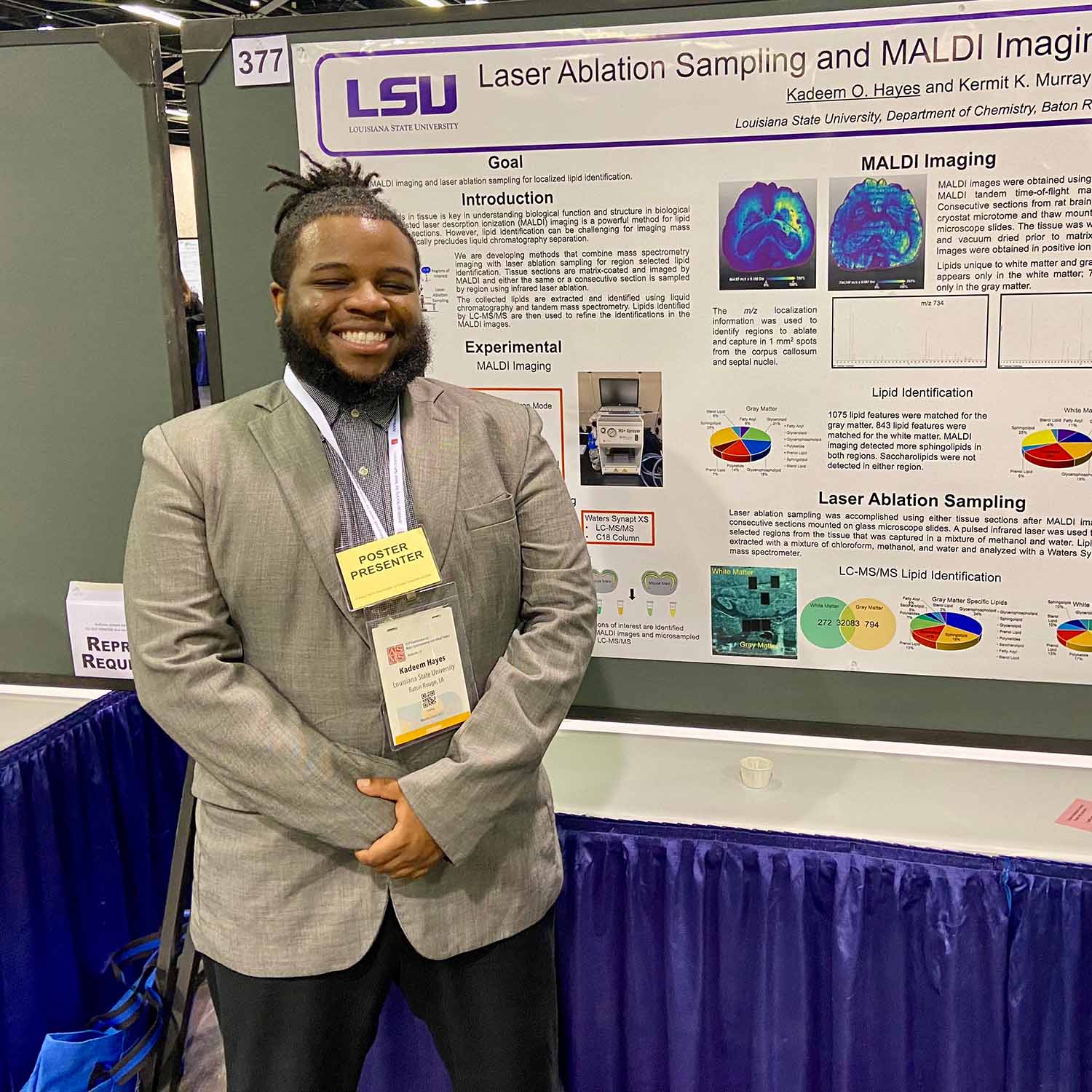
MP 377 Laser Ablation Sampling and MALDI Imaging for Tissue Lipidomics
TP 170 Deep Ultraviolet and Infrared Laser Ablation of Carbonic Anhydrase II
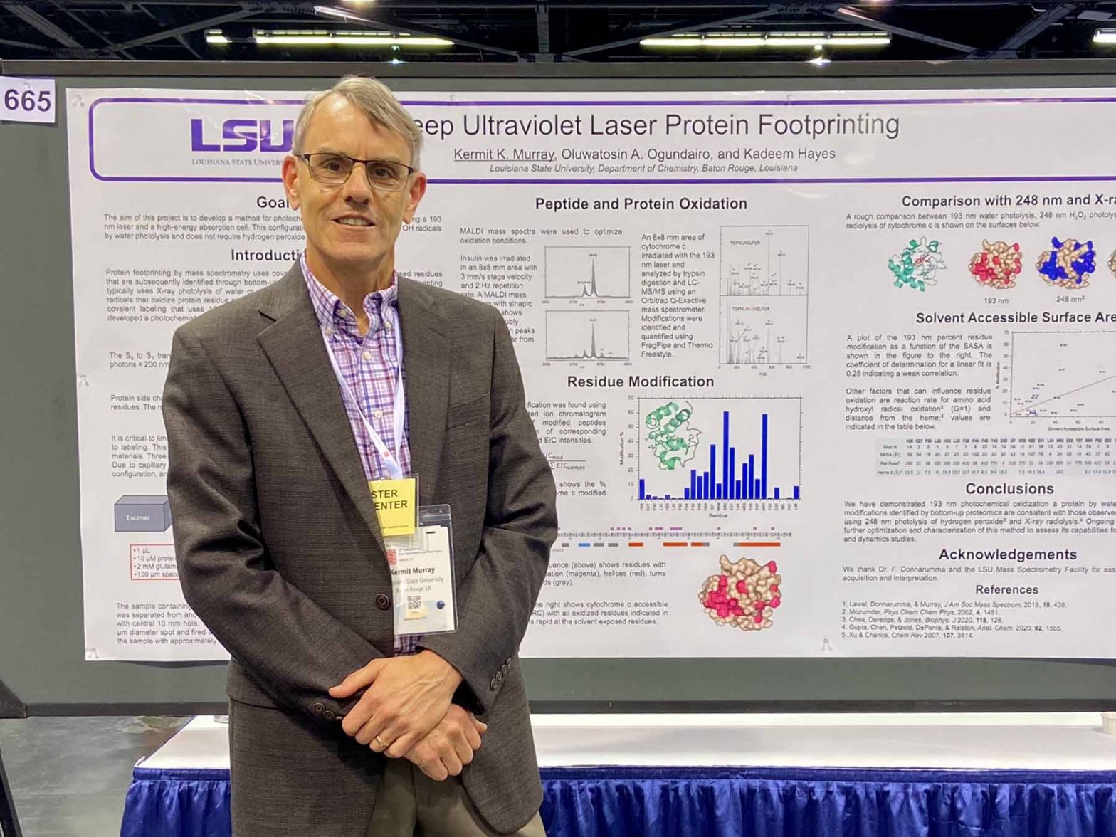
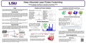
TP 665 Deep Ultraviolet Laser Protein Footprinting
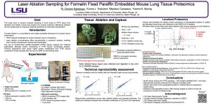
WP 750 Laser Ablation Sampling for Formalin Fixed Paraffin Embedded Mouse Lung Tissue Proteomics
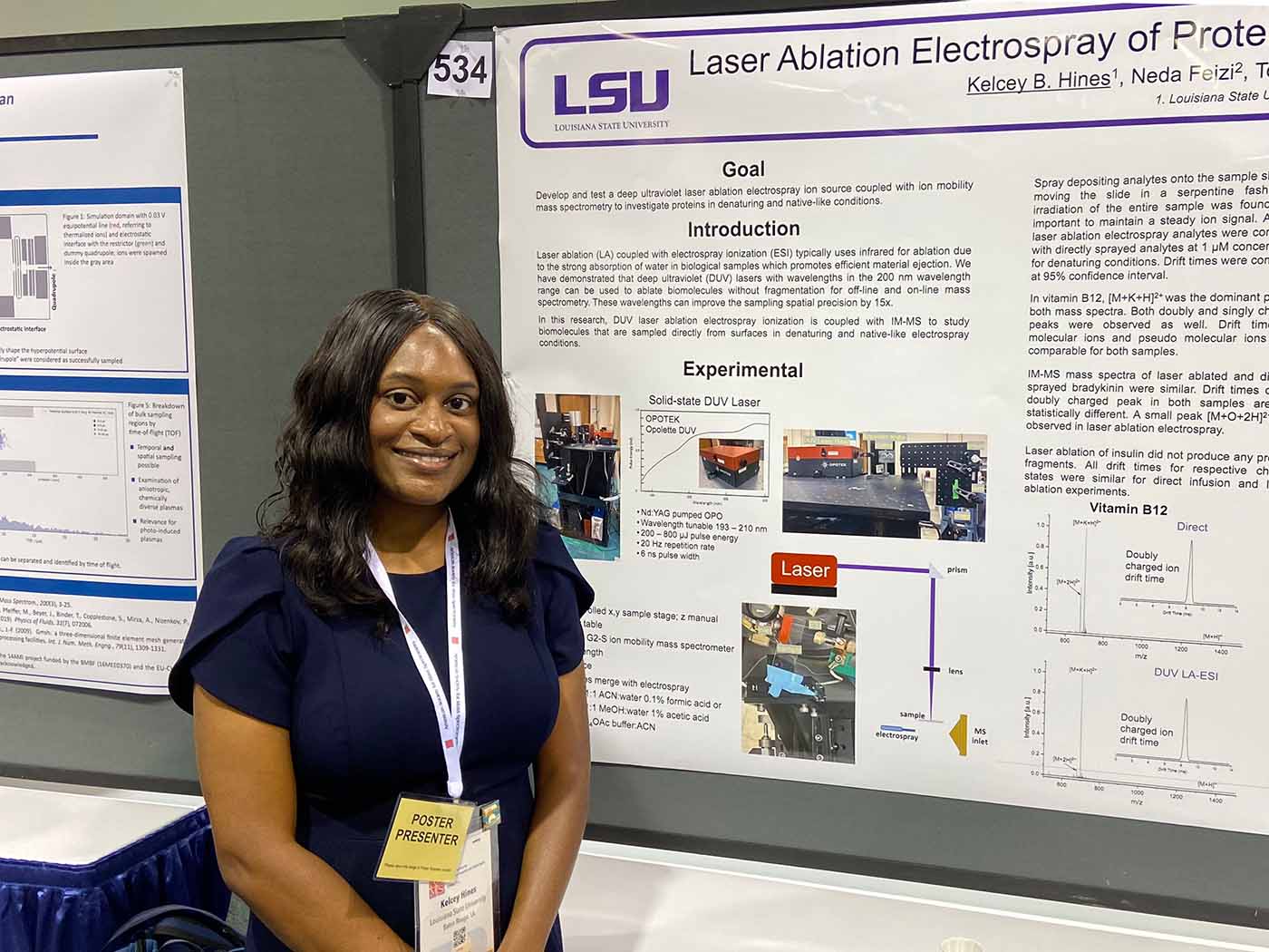
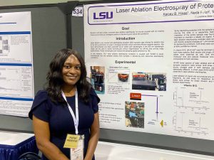
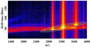
ThP 534 Laser Ablation Electrospray of Proteins with Ion Mobility Mass Spectrometry


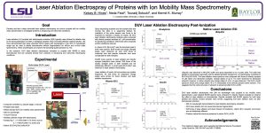


Taylar Bell, Kadeem Hayes, and Kermit K Murray
TP 382 Photochemical Tissue Pre-processing for Matrix-Assisted Laser Desorption Ionization Imaging of Lipids

TP 665 Deep Ultraviolet Laser Protein Footprinting
Kermit K. Murray*; Oluwatosin A. Ogundairo; Kadeem Hayes
Louisiana State University, Baton Rouge, LA
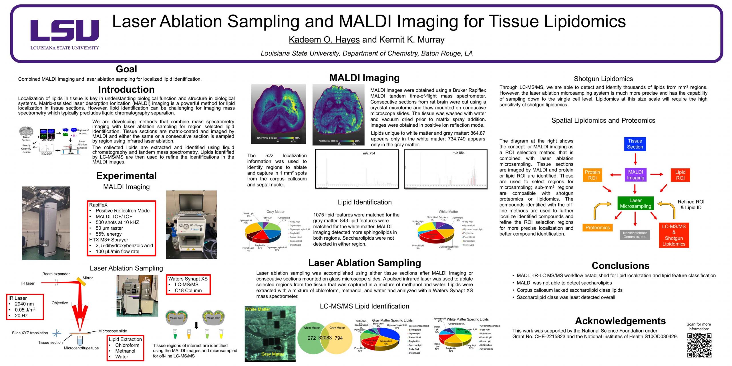
MP 377 Laser Ablation Sampling and MALDI Imaging for Tissue Lipidomics
Kadeem O. Hayes and Kermit K. Murray
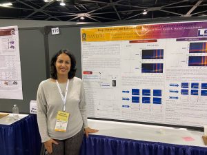
Deep Ultraviolet and Infrared Laser Ablation of Carbonic Anhydrase II
Neda Feizi1; Kadeem O Hayes2; Blessing Chisom Egbejiogu2; Kelcey B. Hines2; Kermit K. Murray2; Touradj Solouki1
1Baylor University, Waco, TX; 2Louisiana State University, Baton Rouge, LA
Introduction
Integrating ion mobility-mass spectrometry (IM-MS) with laser ablation (LA) holds great appeal for acquiring spatially resolved details on protein conformations. Previously, we showed that protein conformation remained intact after LA. However, the potential for laser-induced unfolded proteins to refold back into native-like structures in the capture-solvent remains unresolved. In contrast to typical proteins, denatured bovine carbonic anhydrase II (CA) does not revert to its original conformation in native capture-solvent; instead, it aggregates. This characteristic makes CA particularly well-suited for investigating conformational changes in native surface mass spectrometry (NSMS). Here, we employ NSMS to explore protein unfolding and refolding events, enabling us to examine conformational stabilities during infrared (IR) and deep UV (DUV) laser ablation experiments.
Methods
All solvents and chemicals were purchased from commercial sources and used without further purification. CA stock solution was prepared at 750 µM concentration in native solution of 200 mM ammonium acetate (NH4OAc) adjusted to a pH of 7.3 ± 0.1. Four microliters of the stock solution was deposited on the quartz microscope slide and air-dried. Either a 193 nm ArF excimer laser or 2.94 µm OPO laser was used at a repetition rate of 20 Hz to ablate CA from the surface. All laser ablation experiments were performed in transmission geometry with capturing in 20 µL of native solution. Native IM-MS and broadband collision-induced unfolding (CIU) data were acquired using a Waters Synapt G2-S instrument (Milford, MA).
Preliminary Data
Native mass spectrometry was utilized to probe the charge states and CIU pathways of CA. After deposition and drying on a glass microscopic slide, CA was either washed with native solution or ablated with IR or DUV lasers into the native solution. To minimize variations in mass spectra, including observed charge states and peak shapes, all ESI emitters were inspected with SEM before acquiring mass spectra and only emitters with tip diameters between 600 ± 100 nm were utilized. Mass spectra from triplicate washed and laser-ablated CA showed analogous patterns and charge state distributions (+8 to +10). Moreover, the statistical t-test indicated that the average charge states and peak areas of the washed and laser-ablated CA were not significantly different. The broadband protein ion unfolding experiments were performed without quadrupole ion isolation of specific charge states. Ion collisional activation was achieved in the pre-IM trap cell by establishing a potential difference from 10 to 100 V in 5 V increments. CIU plots showed that the number of unfolding intermediates and the onset voltages for each unfolding transition of washed and laser-ablated CA were similar. The findings from complementary MALDI and bottom-up analyses of native, refolded, and aggregated CA corroborate these observations. The presented work suggests that the native-like conformation of CA remains unchanged after DUV and IR laser ablation; therefore, laser ablation can be used for NSMS.
Novel Aspect
Carbonic anhydrase II serves as a unique probe, enabling the investigation of structural stability during IR and DUV laser ablation.
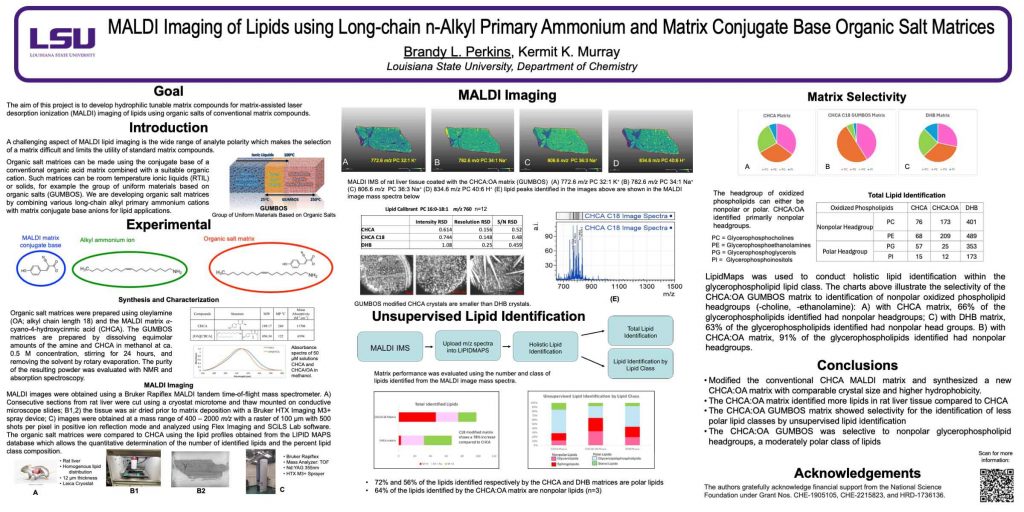
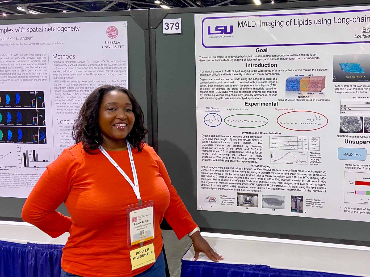
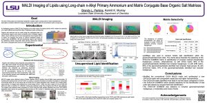
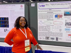
WP 379 MALDI Imaging of Lipids using Long-chain n-Alkyl Primary Ammonium and Conventional Matrix Conjugate Base Organic Salt Matrices
Brandy L. Perkins and Kermit K. Murray
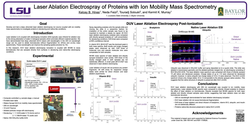
ThP 534 Laser Ablation Electrospray of Proteins with Ion Mobility Mass Spectrometry