K.K. Murray, S. Ghorai, C.A. Seneviratne, Tip Enhanced Laser Ablation Sample Transfer for Mass Spectrometry, MRS Proc. 1754 (2015) mrsf14–1754–pp08–04. doi:10.1557/opl.2015.286.
Abstract

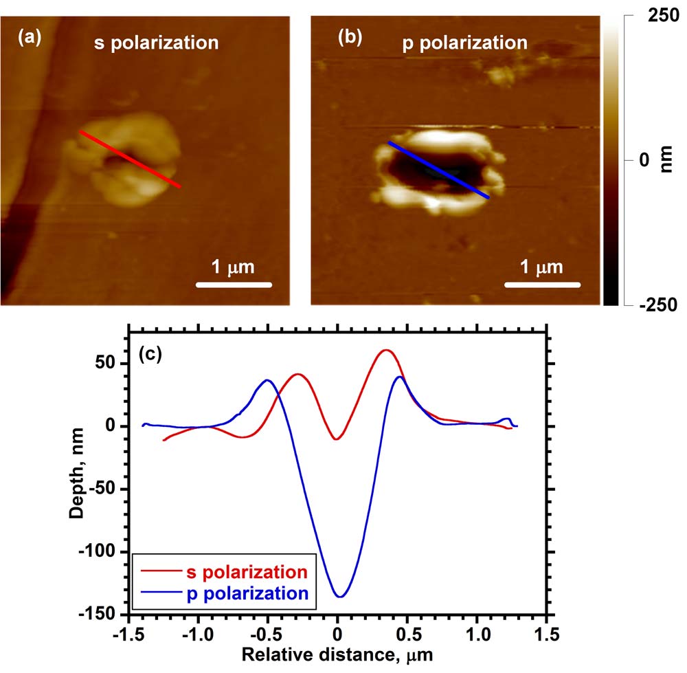
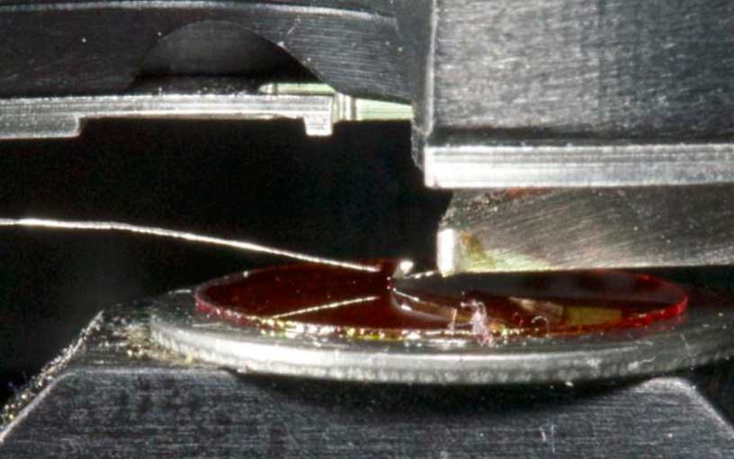
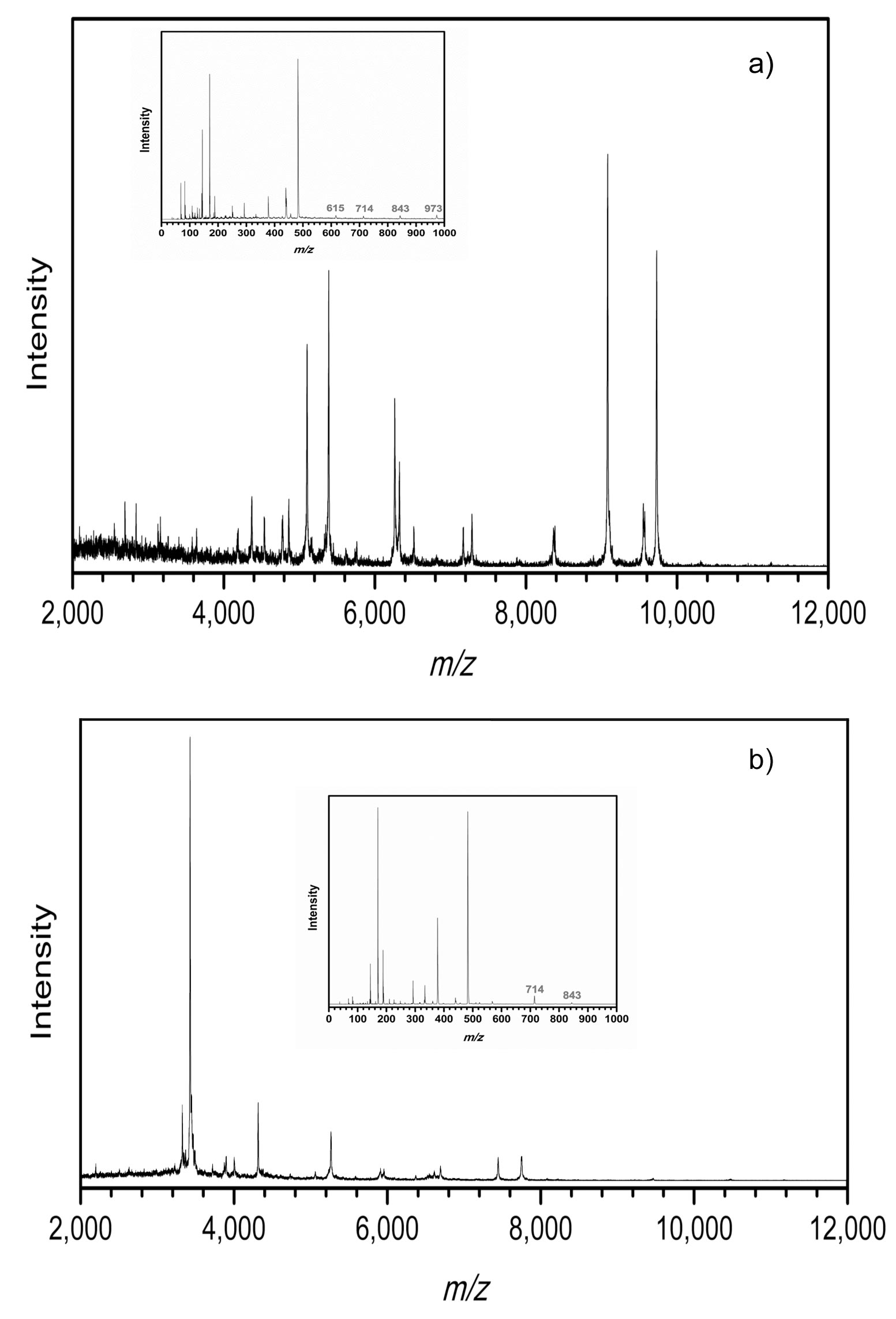
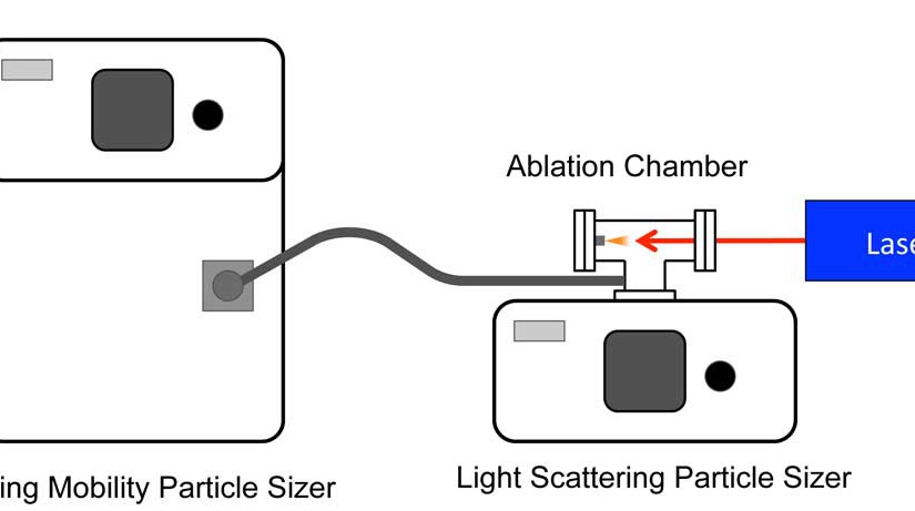
Mass spectrometry is one of the primary analysis techniques for biological analysis but there are technological barriers in sampling scale that must be overcome for it to be used to its full potential on the size scale of single cells. Current mass spectrometry imaging methods are limited in spatial resolution when analyzing large biomolecules. The goal of this project is to use atomic force microscope (AFM) tip enhanced laser ablation to remove material from cells and tissue and capture it for subsequent mass spectrometry analysis. The laser ablation sample transfer system uses an AFM stage to hold the metal-coated tip at a distance of approximately 10 nm from a sample surface. The metal tip acts as an antenna for the electromagnetic radiation and enables the ablation of the sample with a spot size much smaller than a laser focused with a conventional lens system. A pulsed nanosecond UV or visible wavelength laser is focused onto the gold-coated silicon tip at an angle nearly parallel with the surface, which results in the removal of material from a spot between 500 nm and 1 µm in diameter and 200 and 500 nm deep. This corresponds to a few picograms of ablated material, which can be captured on a metal surface for MALDI analysis. We have used this approach to transfer small peptides and proteins from a thin film for analysis by mass spectrometry as a first step toward high spatial resolution imaging.


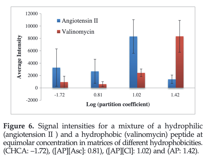


")