Particle size measurement from infrared laser ablation of tissue

F. Cao, F. Donnarumma, K.K. Murray, “Particle size measurement from infrared laser ablation of tissue,” Analyst. 141 (2016) 183โ€“190. doi:10.1039/C5AN01765C.

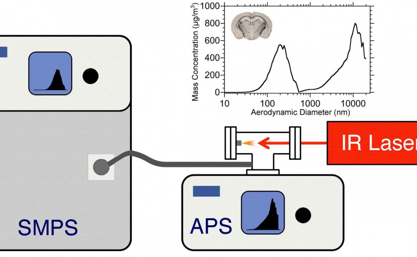
Abstract: The concentration and size distribution were measured for particles ablated from tissue sections using an infrared optical parametric oscillator laser system. A scanning mobility particle sizer and light scattering particle sizer were used in parallel to realize a particle sizing range from 10 nm to 20 ฮผm. Tissue sections from rat brain and lung ranging in thickness between 10 and 50 ฮผm were mounted on microscope slides and irradiated with nanosecond laser pulses at 3 ฮผm wavelength and fluences between 7 and 21 kJ mโˆ’2 in reflection geometry. The particle size distributions were characterized by a bimodal distribution with a large number of particles 100 nm in diameter and below and a large mass contribution from particles greater than 1 ฮผm in diameter. The large particle contribution dominated the ablated particle mass at high laser fluence. The tissue type, thickness, and water content did not have a significant effect on the particle size distributions. The implications of these results for laser ablation sampling and mass spectrometry imaging under ambient conditions are discussed.

Cao Analyst 2016
Particle size measurement from infrared laser ablation of tissue, Analyst. 141 (2016) 183

ASMS 2016: Laser Ablation Sample Transfer with Vacuum Capture for Forensic Evidence Sampling

 Laser Ablation Sample Transfer with Vacuum Capture for Forensic Evidence Sampling
Laser Ablation Sample Transfer with Vacuum Capture for Forensic Evidence Sampling, F. Donnarumma, E. Camp, and K. K. Murray; ASMS 2016, San Antonio

Ablation of antibacterial cream

Fingerprint ablation

Ablation and capture with different filters

Ablation on Textile

High Resolution Laser Mass Spectrometry Bioimaging

K. K. Murray, , C. A. Seneviratne, and S. Ghorai, “High Resolution Laser Mass Spectrometry Bioimaging” Methods 104 (2016) 118โ€“126; doi:10.1016/j.ymeth.2016.03.002

Mass spectrometry imaging (MSI) was introduced more than five decades ago with secondary ion mass spectrometry (SIMS) and a decade later with laser desorption/ionization (LDI) mass spectrometry (MS). Large biomolecule imaging by matrix-assisted laser desorption/ionization (MALDI) was developed in the 1990s and ambient laser MS a decade ago. Although SIMS has been capable of imaging with a moderate mass range at sub-micrometer lateral resolution from its inception, laser MS requires additional effort to achieve a lateral resolution of 10 lm or below which is required to image at the size scale of single mammalian cells. This review covers untargeted large biomolecule MSI using lasers for desorption/ionization or laser desorption and post-ionization. These methods include laser microprobe (LDI) MSI, MALDI MSI, laser ambient and atmospheric pressure MSI, and near-field laser ablation MS. Novel approaches to improving lateral resolution are discussed, including oversampling, beam shaping, transmission geometry, reflective and through-hole objectives, microscope mode, and near-field optics.

https://kermitmurray.com/documents/murray_2016.pdf

Laser Ablation Sample Transfer for Localized LC-MS/MS Proteomic Analysis of Tissue

F. Donnarumma, F. Cao, K.K. Murray, Laser Ablation with Vacuum Capture for MALDI Mass Spectrometry of Tissue, J. Am. Soc. Mass Spectrom. 27 (2016) 108โ€“116. doi:10.1007/s13361-015-1249-0.

Abstract: We have developed a laser ablation sampling technique for matrix-assisted laser desorption ionization (MALDI) mass spectrometry and tandem mass spectrometry (MS/MS) analyses of in-situ digested tissue proteins. Infrared laser ablation was used to remove biomolecules from tissue sections for collection by vacuum capture and analysis by MALDI. Ablation and transfer of compounds from tissue removes biomolecules from the tissue and allows further analysis of the collected material to facilitate their identification. Laser ablated material was captured in a vacuum aspirated pipette-tip packed with C18 stationary phase and the captured material was dissolved, eluted, and analyzed by MALDI. Rat brain and lung tissue sections 10ย ฮผm thick were processed by in-situ trypsin digestion after lipid and salt removal. The tryptic peptides were ablated with a focused mid-infrared laser, vacuum captured, and eluted with an acetonitrile/water mixture. Eluted components were deposited on a MALDI target and mixed with matrix for mass spectrometry analysis. Initial experiments were conducted with peptide and protein standards for evaluation of transfer efficiency: a transfer efficiency of 16% was obtained using seven different standards. Laser ablation vacuum capture was applied to freshly digested tissue sections and compared with sections processed with conventional MALDI imaging. A greater signal intensity and lower background was observed in comparison with the conventional MALDI analysis. Tandem time-of-flight MALDI mass spectrometry was used for compound identification in the tissue. Graphical Abstract แ…Ÿ.

Laser Ablation Sample Transfer for Localized LC-MS/MS Proteomic Analysis of Tissue. J. Mass Spectrom. 2016, 51, 261
J. Mass Spectrom. 2016, 51, 261โ€“268.
Laser Ablation with Vacuum Capture for MALDI Mass Spectrometry of Tissue