F. Cao, F. Donnarumma, K.K. Murray, Wavelength-Dependent Tip-Enhanced Laser Ablation of Organic Dyes, Journal of Physical Chemistry C, 124 (2020) 1918-1922; doi: 10.1021/acs.jpcc.9b08081
Abstract
Wavelength-Dependent Tip-Enhanced Laser Ablation of Organic Dyes
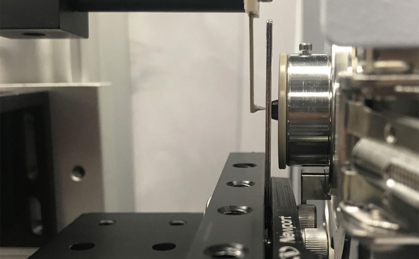
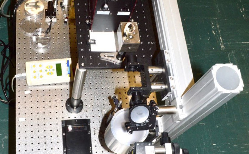
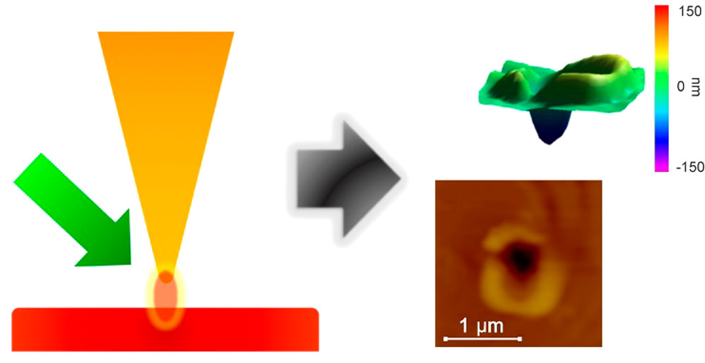
The wavelength dependence of atomic force microscope apertureless tip-enhanced laser ablation was studied using a series of organic dyes to assess the effect of surface optical absorption. An optical parametric oscillator laser with a tunable wavelength range of 410-2100 nm was used to irradiate a gold-coated atomic force microscope tip which was held 15 nm above the surface of a dye thin film vibrating in tapping mode to generate tip-enhanced laser ablation. Crater formation was investigated for rhodamine B, methylene blue, and IR 797 dye thin films which have absorption maxima near 550, 650, and 700 nm, respectively. Crater formation was not observed for wavelengths greater than 700 nm for any of the dyes. Below 700 nm, the crater size was greatest near 500 nm for all dyes and decreased from 500 to 700 nm, and no ablation was observed at longer wavelengths. The crater size did not correlate with the solution or thin film optical absorption of the dyes. The mechanism for tip-enhanced laser ablation is postulated to be either ballistic ejection of gold atoms or direct heat transfer from the tip to the surface.