Research group of Kermit Murray at Louisiana State University where we use lasers for sampling and imaging and study the chemistry and physics of laser ablation.
K.K. Murray, Mass spectrometry and Web 2.0, J. Mass Spectrom. 42 (2007) 1263–1271. doi:10.1002/jms.1315.
Abstract
The term Web 2.0 is a convenient shorthand for a new era in the Internet in which users themselves are both generating and modifying existing web content. Several types of tools can be used. With social bookmarking, users assign a keyword to a web resource and the collection of the keyword ‘tags’ from multiple users form the classification of these resources. Blogs are a form of diary or news report published on the web in reverse chronological order and are a popular form of information sharing. A wiki is a website that can be edited using a web browser and can be used for collaborative creation of information on the site. This article is a tutorial that describes how these new ways of creating, modifying, and sharing information on the Web are being used for on-line mass spectrometry resources.
Presented at the 55th ASMS Conference on Mass Spectrometry, June 4, 2007, Indianapolis, Indiana
Mass Spectrometry on Wikipedia: Open Source and Peer Review, Mass Spectrometry on Wikipedia: Open Source and Peer Review
Goal
Develop a procedure for peer review of Wikipedia articles on mass spectrometry.
Introduction
Wikipedia is an on-line encyclopedia that anyone can edit. The site is maintained by hundreds of users who create, edit and organize articles, oversee quality and consistency, moderate disputes, and guard against abuse. Anyone can contribute as much or as little as he or she desires. On the English language Wikipedia in early 2007, there were 1.4 M articles being edited by more than 150,000 users who were creating about 2000 new articles each day. Of these articles, 70 were in the category “Mass Spectrometry.”
Why is Wikipedia Important?
The public face of mass spectrometry is determined by the information that can be found most easily on the web.
Content Development Models
In April 2007, the top ten hits for “mass spectrometry” on the search engine Google were
Wikipedia
ASMS (What is MS?)
ASMS
Michigan State University
University of Arizona
University of Leeds
University of Illinois, Chicago
Scripps Institute
Ionsource.com
Imass.com
and on Yahoo
Wikipedia
I-mass.com
Basepeak.com
Encyclopaedia Britannica
ASMS
Michigan State University
Wikipedia
GenomicGlossaries.com
Mass Spectrometry Blog
University of New Mexico
Wikipedia is the #1 hit for “Mass Spectrometry” on the top five search pages
Google
Yahoo
Ask.com
MSN Live Search
Altavista
Wikipedia is the way that the public learns about mass spectrometry on the web yet the information is not subject to peer review and it is not developed in conjunction with any mass spectrometry organization.
Content Development Models
Three basic content development models are presented with their advantages and disadvantages. Here, a Wikipedian is someone who understands the workings of Wikipedia and can create and edit content and work with Editors, Bureaucrats and Administrators. A mass spectrometrist is a person who has or is working toward an advanced degree in mass spectrometry or has the equivalent work experience.
Open Model
The Open Model is in effect today. Wikipedians, who may also be mass spectrometrists, openly edit mass spectrometry content on Wikipedia, drawing on mass spectrometry literature and web-based resources.
Wikipedia Open Model
Advantages and Disadvantages
The advantage of the open model is that it is already in place. There are mass spectrometry entries on Wikipedia and anyone can add content within the bounds of the Wikipedia community. Wikipedians with a knowledge of mass spectrometry, chemistry, physics and other areas provide an ad hoc peer review.
The disadvantages are the time required to learn the Wikipedia system to effectively generate content and the fact that the site tends to favor the persistent and tactically adroit over the subject expert. Further, content generated on Wikipedia is not credited to the student or academic and copyright is not retained.
Closed Model
In the closed model, content is generated by mass spectrometrists and in many cases subject to peer review. For example, the ASMS “What is Mass Spectrometry?” web page. Another example is mass spectrometry journals, all of which are on line and some of these are openly available.
Advantages and Disadvantages
The advantage of the closed model is that the content is created by experts and peer reviewed. The disadvantage is that the information is not generally available and is often presented at a level not suited to a general audience. Some excellent works have been generated in recent years, such as the documents created by the ASMS Education Committee, Michael A Grayson’s Measuring Mass: From Positive Rays to Proteins, and a wide range of excellent mass spectrometry textbooks. However, only a few of these works are openly available. Consider a high school student writing a report on mass spectrometry. Where will that student obtain the information for that report?
The Protected Fork Model
The material in Wikipedia is not copyrighted and it can therefore be “forked” – all or part of the material can be removed and independently modified as long as the derivative material is also made available with the same unrestricted license. This is the kind of reciprocal license principle that is applied to open source software.
A Wikipedia fork must be available by unrestricted license, but it need not be openly edited: the fork can be protected. Under this model, mass spectrometry content is taken from Wikipedia under the terms of the GNU Free Documentation License. This “Mass Spectrometry Wiki” can only be modified by a set of approved mass spectrometry editors whose work is reviewed by their peers. Non-wikipedian mass spectrometrists can participate without the need to learn the rules and language of generating Wikipedia content. Wikipedian mass spectrometrists can help to synchronize (or at least harmonize) the content between the two wikis.
Wikipedia protected fork model
Advantages and Disadvantages
The advantage of the Protected Fork Model is that it has the potential to bring in experts on a subject who may not otherwise be inclined to participate directly in Wikipedia. It offers the possibility of peer review within the Wikipedia construct. The disadvantage is the difficulty in synchronizing information (in both directions) and the need for experienced Wikipedians who are also experienced mass spectrometrists.
Conclusions
Wikipedia’s presentation of mass spectrometry has become a significant part of the way the public perceives the field. The mass spectrometry community should be aware of this fact and avenues for participation in Wikipedia should be encouraged and facilitated.
J. Dong, Y. H. Rezenom, and K. K. Murray, “Aerosol Desorption Electrospray Ionization,” Presented at the 55th ASMS Conference on Mass Spectrometry, June 4, 2007, Indianapolis, Indiana, Ambient Ionization I, MP 006.
M.W. Little, K.K. Murray, “Two-Laser Mid-Infrared and Ultraviolet Matrix-Assisted Laser Desorption/Ionization,” Int. J. Mass Spectrom.261 (2007) 140–145. doi:10.1016/j.ijms.2006.08.010.
Abstract
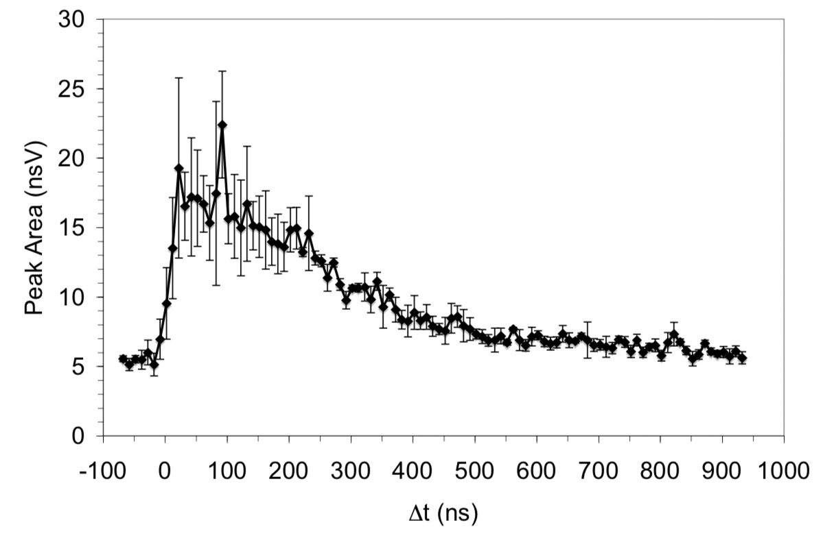
Integrated ion signal for the NO loss fragment of 4-nitroaniline plotted as a function of delay time from −70 to 950 ns in 10 ns increments. Error bars represent one standard deviation.
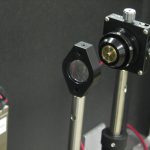
Matrix-assisted laser desorption/ionization (MALDI) was performed using two-pulsed lasers with wavelengths in the infrared and ultraviolet regions. A 2.94 μm pulsed optical parametric oscillator laser system and a 337 nm pulsed nitrogen laser irradiated the same spot on the sample target. Sinapinic acid (SA), 2,5-dihydroxybenzoic acid (DHB), α-cyano-4-hydroxycinnamic acid (CCA), and 4-nitroaniline (NA) were used as matrices, and bovine insulin and cytochrome C were used as analytes. The laser energy was adjusted so that one-laser MALDI and LDI was at a minimum and the matrix and analyte ion signal was enhanced when the two lasers were fired together. Two-laser LDI was observed with SA, DHB, and NA matrices and two-laser MALDI was observed with SA and DHB. Plots of ion signal as a function of delay between the IR and UV lasers show two-laser signal from 0 ns up to a delay of 500 ns when the IR laser is fired before the UV laser. The results are interpreted in terms of IR laser heating of the target that leads to an enhancement in UV LDI and MALDI.
M.W. Little, J. Laboy, K.K. Murray, Wavelength dependence of soft infrared laser desorption and ionization, J. Phys. Chem. C.111 (2007) 1412–1416. doi:10.1021/jp063154v.
Abstract
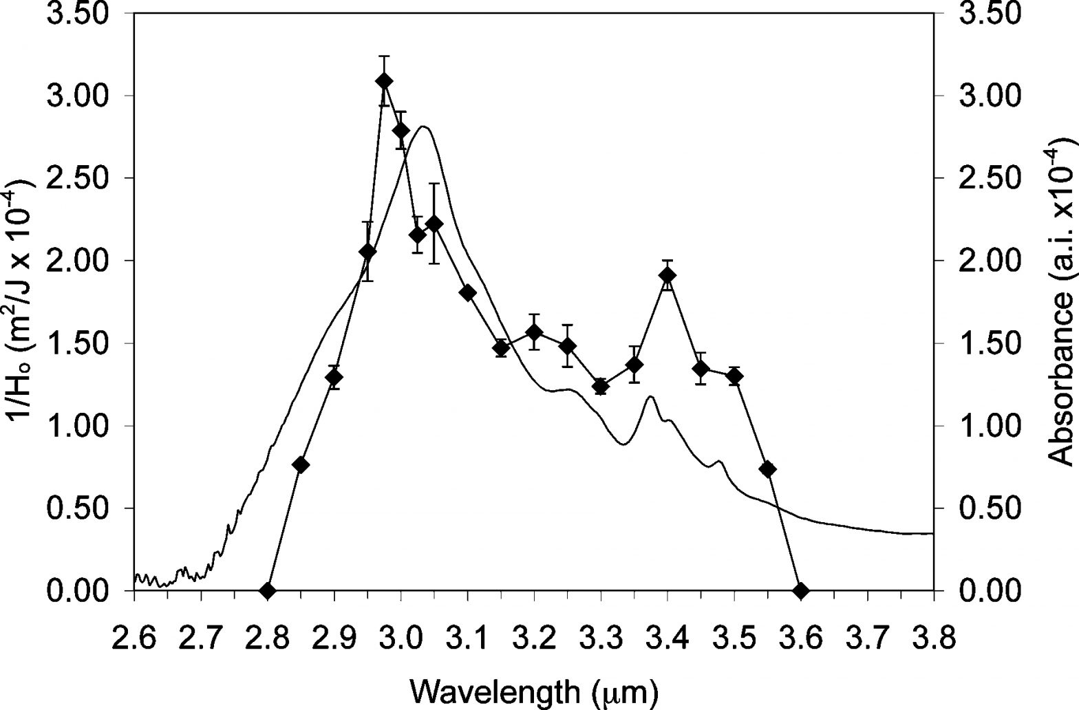
Inverse threshold fluence for insulin ion formation plotted as a function of wavelength (solid line and solid circles with one standard deviation error bar) overlaid with the IR absorption spectrum of an insulin thin film (solid line, no data symbols).
Protonated insulin molecules were formed by soft IR laser desorption ionization of a thin film of the protein on a silicon surface. Time-of-flight mass spectra were recorded at wavelengths between 2.8 and 3.6 µm and the efficiency of ionization was compared to the IR absorption of the protein thin film. Ionization efficiency was quantified by recording the minimum laser energy per unit area required to produce a detectable ion signal (threshold fluence). The ionization efficiency tracks the IR absorption spectrum of insulin between 2.6 and 3.8 µm in the region of OH, NH, and CH stretch absorption. The lowest threshold fluence and therefore the most efficient ionization was nearly coincident with the OH stretch absorption of insulin near 3.0 µm. An additional local maximum in ionization efficiency was observed at 3.4 µm, coincident with the CH stretch vibrational absorption. Comparison of the ionization efficiency with the IR absorption indicates that the protein and not the residual solvent is absorbing the laser energy. Scanning electron microscopy images of the bovine insulin thin films on silicon after laser irradiation show melting and indications of explosive boiling. Ionization occurs through the sacrifice of some of the protein molecules that absorb the laser energy and act as an intrinsic matrix.
S.N. Jackson, J.-K. Kim, J.L. Laboy, K.K. Murray, Particle formation by infrared laser ablation of glycerol: implications for ion formation, Rapid Commun. Mass Spectrom.20 (2006) 1299–1304. doi:10.1002/rcm.2443.
Abstract
Particle sized distribution weighted by (a) count and (b) mass from ablation of glycerol at a wavelength of 2.95 µm with a fluence of 4500 J/m2.
The quantity and size distribution of micrometer-sized particles ejected from thin films of glycerol were measured using light scattering particle sizing. Thin glycerol films were irradiated at atmospheric pressure with an infrared optical parametric oscillator at wavelengths between 2.95 and 3.1 µm. Particulate material resulting from the ablation was sampled directly into a particle-sizing instrument and particles with diameters greater than 500 nm were detected and sized by light scattering. The fluence threshold for particle formation was between 2000 and 3000 J/m2 for all laser wavelengths. At threshold, fewer than 100 particles/cm3 were detected and this value increased to several thousand particles/cm3 at twice the threshold fluence. The average size of the coarse particles ranged from 900 nm to 1.6 µm at threshold and decreased by 10-20% at twice the threshold fluence. The coarse particle formation observations were compared with ion formation behavior in matrix-assisted laser desorption ionization and interpreted in terms of a photomechanical mechanism for material ablation and ion formation.
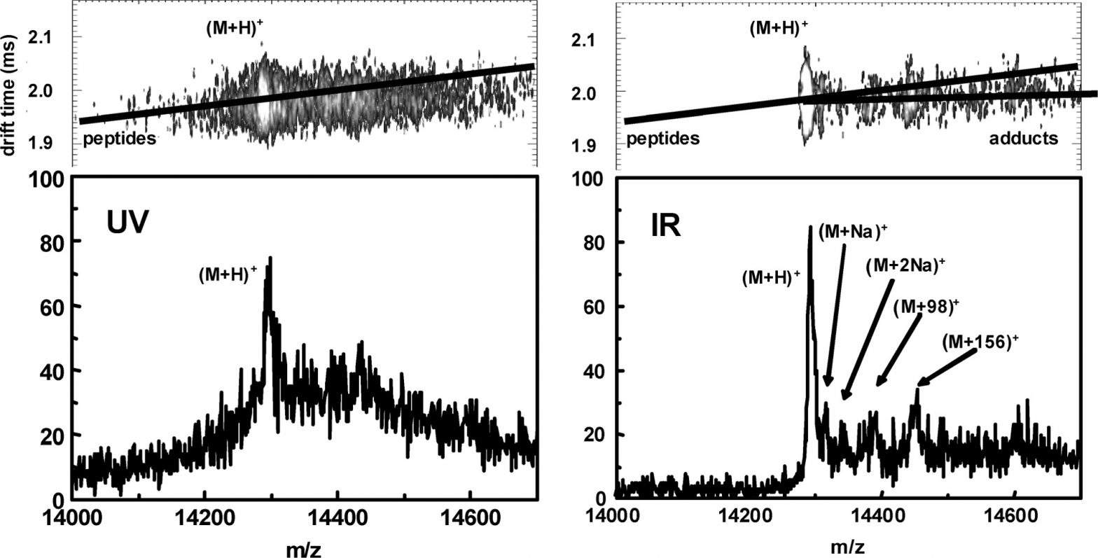
A.S. Woods, M.V. Ugarov, S.N. Jackson, T. Egan, H.-Y.J. Wang, K.K. Murray, J. A. Schultz, “IR-MALDI-LDI combined with ion mobility orthogonal time-of-flight mass spectrometry,” J. Proteome Res.5 (2006) 1484–1487. doi:10.1021/pr060055l.
Abstract
Woods, Ugarov, Jackson, Egan, Wang, Murray, & Schultz, IR-MALDI-LDI combined with ion mobility orthogonal time-of-flight mass spectrometry, J. Proteome Res. 5 (2006) 1484.
Most MALDI instrumentation uses UV lasers. We have designed a MALDI−IM−oTOF−MS which employs both a Nd:YAG laser pumped optical parametric oscillator (OPOTEK, λ = 2.8−3.2 μm at 20 Hz) to perform IR−LDI or IR−MALDI and a Nd:YLF laser (Crystalaser, λ = 249 nm at 200 Hz) for the UV. Ion mobility (IM) gives a fast separation and analysis of biomolecules from complex mixtures in which ions of similar chemical type fall along well-defined “trend lines”. Our data shows that ion mobility allows multiply charged monomers and multimers to be resolved; thus, yielding pure spectra of the singly charged protein ion which are virtually devoid of chemical noise. In addition, we have demonstrated that IR−LDI produced similar results as IR−MALDI for the direct tissue analysis of phospholipids from rat brain.
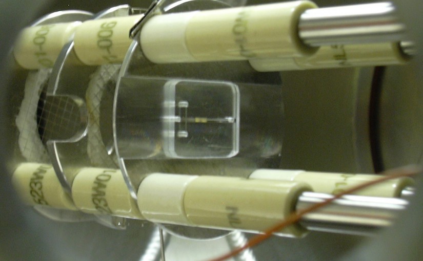
Y. Xu, M.W. Little, K.K. Murray, “Interfacing capillary gel microfluidic chips with infrared laser desorption mass spectrometry,” J. Am. Soc. Mass Spectrom.17 (2006) 469–474. doi:10.1016/j.jasms.2005.12.003.
Abstract
Capillary gel microfluidic chip interfaced to laser desorption/ionization (LDI) mass spectrometry with a time-of-flight mass analyzer.
We report on the fabrication and performance of a gel microfluidic chip interfaced to laser desorption/ionization (LDI) mass spectrometry with a time-of-flight mass analyzer. The chip was fabricated from poly(methylmethacrylate) with a poly(dimethyl siloxane) cover. Sodium dodecyl sulfate-polyacrylamide gel electrophoresis was performed in the channel of the microfluidic chip. After electrophoresis, the cover was removed and either the PDMS chip or the PMMA cover was mounted in a modified MALDI ion source for analysis. Ions were formed by irradiating the channel with 2.95 µm radiation from a pulsed optical parametric oscillator (OPO), which is coincident with IR absorption by N-H and O-H stretch of the gel components. No matrix was added. The microfluidic chip design allowed a decrease in the volume of material required for analysis over conventional gel slabs, thus enabling improvement in the detection limit to a pmol level, a three orders of magnitude improvement over previous studies in which desorption was achieved from an excised section of a conventional gel.
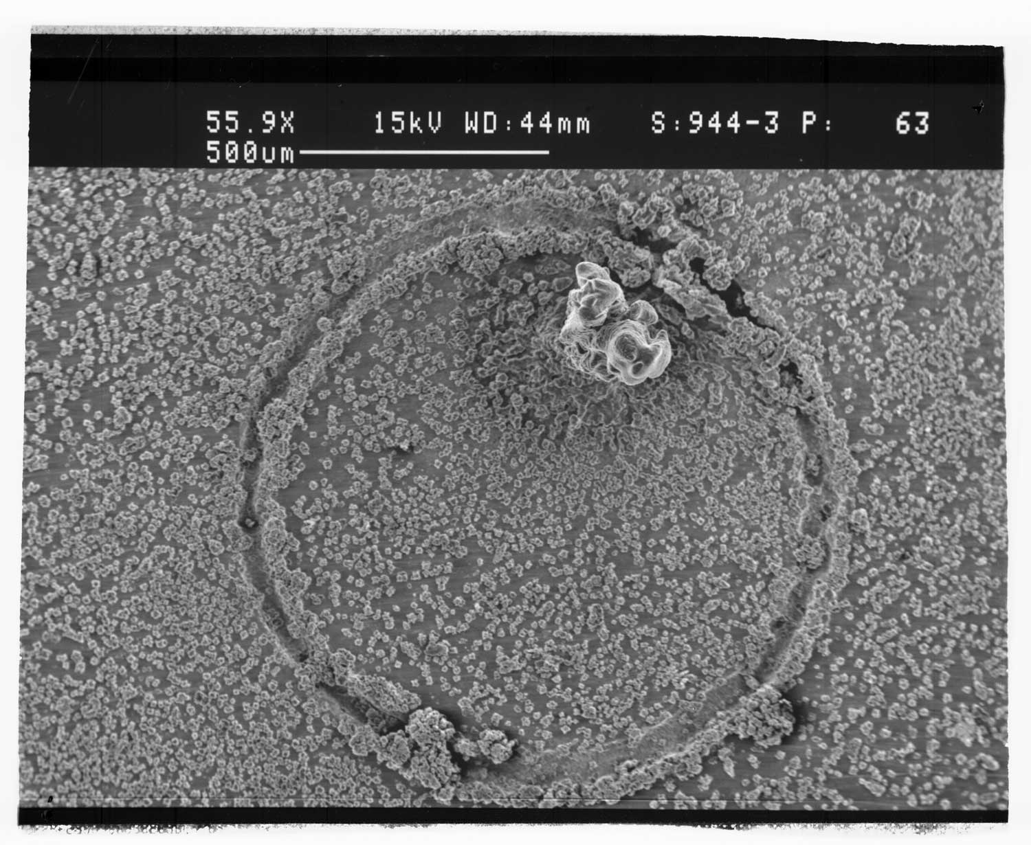
J.-K. Kim, S.N. Jackson, K.K. Murray, Matrix-assisted laser desorption/ionization mass spectrometry of collected bioaerosol particles, Rapid Commun. Mass Spectrom.19 (2005) 1725–1729. doi:10.1002/rcm.1982.
Abstract
Scanning electron microscope image of aerosol deposition on a MALDI target spot
A method was developed for collection and analysis of bioaerosols by matrix-assisted laser desorption/ionization (MALDI) time-of-flight mass spectrometry using a modified Andersen N6 bioaerosol collector. The overall goal of the study was to develop methods for obtaining mass spectra with minimal reagents and treatment steps for potential use in remote collection and analysis systems. Test bioaerosol particles were generated from a nebulized E. coli bacterial suspension and collected on MALDI targets placed in an Andersen N6 single-stage aerosol impactor. The bioaerosols were mixed with matrix either by deposition on a bare target with the matrix solution added later, or by deposition on a target pre-coated with matrix. The matrix compounds ?-cyano-4-hydroxycinnamic acid (CHCA) and sinapic acid (SA) were tested and the SA matrix was found to give the best results in number of peaks, resolution, and signal-to-noise ratio. Deposition of bioaerosol particles onto the matrix pre-coated target did not produce signal in the m/z region above 1000, but the signal could be recovered with the addition of a 1:1 (v/v) acetonitrile/water solvent. Addition of solvent by pipette to the pre-coated targets after particle deposition recovered signal comparable to the dried-droplet sample preparations, whereas solvent sprayed into the impactor recovered fewer peaks. Deposition on pre-coated targets with post-collection solvent addition was superior to deposition on bare target followed by post-collection addition of matrix solution.
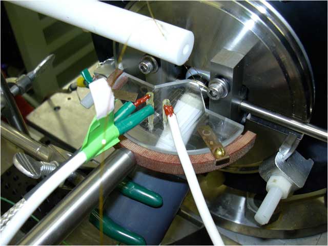
H. Musyimi, J. Guy, D.A. Narcisse, S.A. Soper, K.K. Murray, Direct coupling of polymer-based microchip electrophoresis to online MALDI-MS using a rotating ball inlet, Electrophoresis. 26 (2005) 4703–4710. doi:10.1002/elps.200500317.
Abstract
Rotating ball inlet with capillary electrophoresis microfluidic chip.
We report on the coupling of a polymer-based microfluidic chip to a MALDI-TOF MS using a rotating ball interface. The microfluidic chips were fabricated by micromilling a mold insert into a brass plate, which was then used for replicating polymer microparts via hot embossing. Assembly of the chip was accomplished by thermally annealing a cover slip to the embossed substrate to enclose the channels. The linear separation channel was 50 microm wide, 100 microm deep, and possessed an 8 cm effective length separation channel with a double-T injector (V(inj) = 10 nL). The exit of the separation channel was machined to allow direct contact deposition of effluent onto a specially constructed rotating ball inlet to the mass spectrometer. Matrix addition was accomplished in-line on the surface of the ball. The coupling utilized the ball as the cathode transfer electrode to transport sample into the vacuum for desorption with a 355 nm Nd:YAG laser and analyzed on a TOF mass spectrometer. The ball was cleaned online after every rotation. The ability to couple poly(methylmethacrylate) microchip electrophoresis devices for the separation of peptides and peptide fragments produced from a protein digest with subsequent online MALDI MS detection was demonstrated.


")