In March of 2009, I visited McNeese State University in Lake Charles, Louisiana to give a talk and visit my collaborator Prof. Mark Merchant, “the alligator man”. Mark gave me a tour of his alligator holding facilities on campus.

Mark wanted to get this alligator to open its mouth so that I could get a good photo.

Sure enough, the alligator did open his mouth. Note Chemistry Department Head Ron Darbeau who was on the proper side of the fence during all of this. I was 15 feet away and the gate was open.


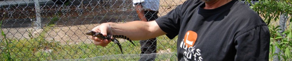
In the next cage over, Mark scooped out a handful of baby alligators.

I call the one on the right “Bitey” – he clamped down on my knuckle when I unwisely offered it to him.