Customer Spotlight – Dr. Kermit Murray – Louisiana State University


Murray Mass Spectrometry Group
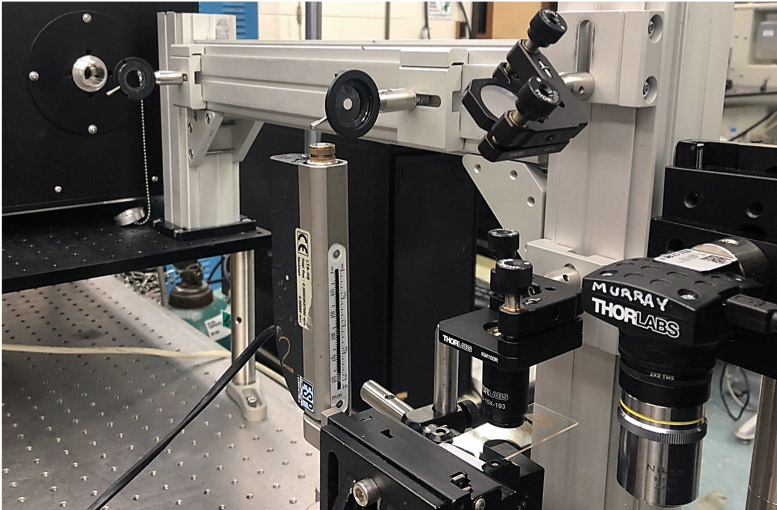
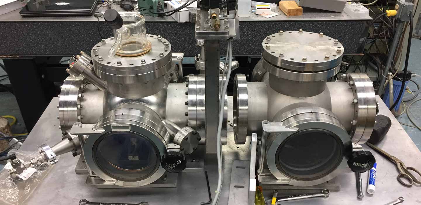
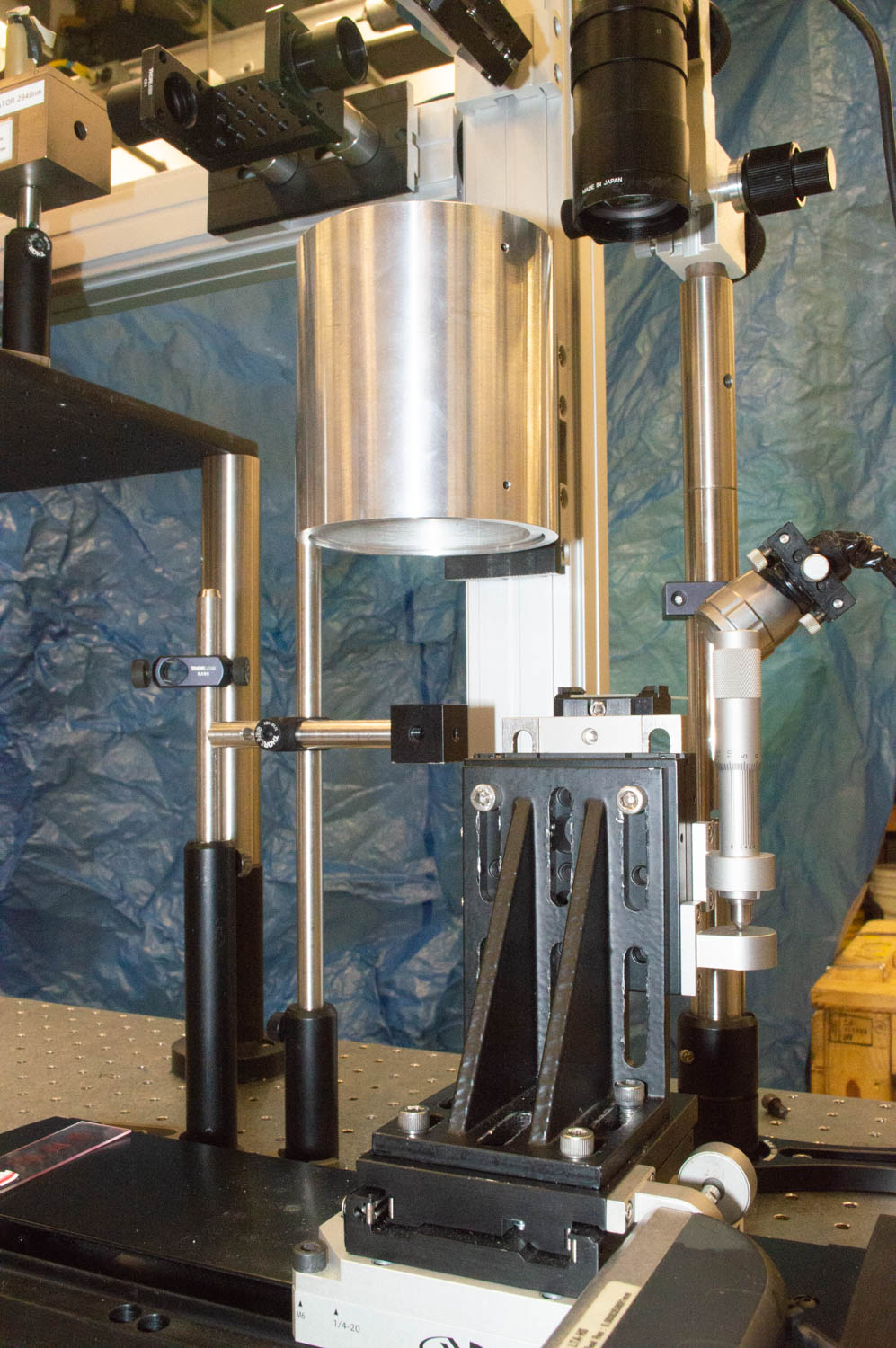
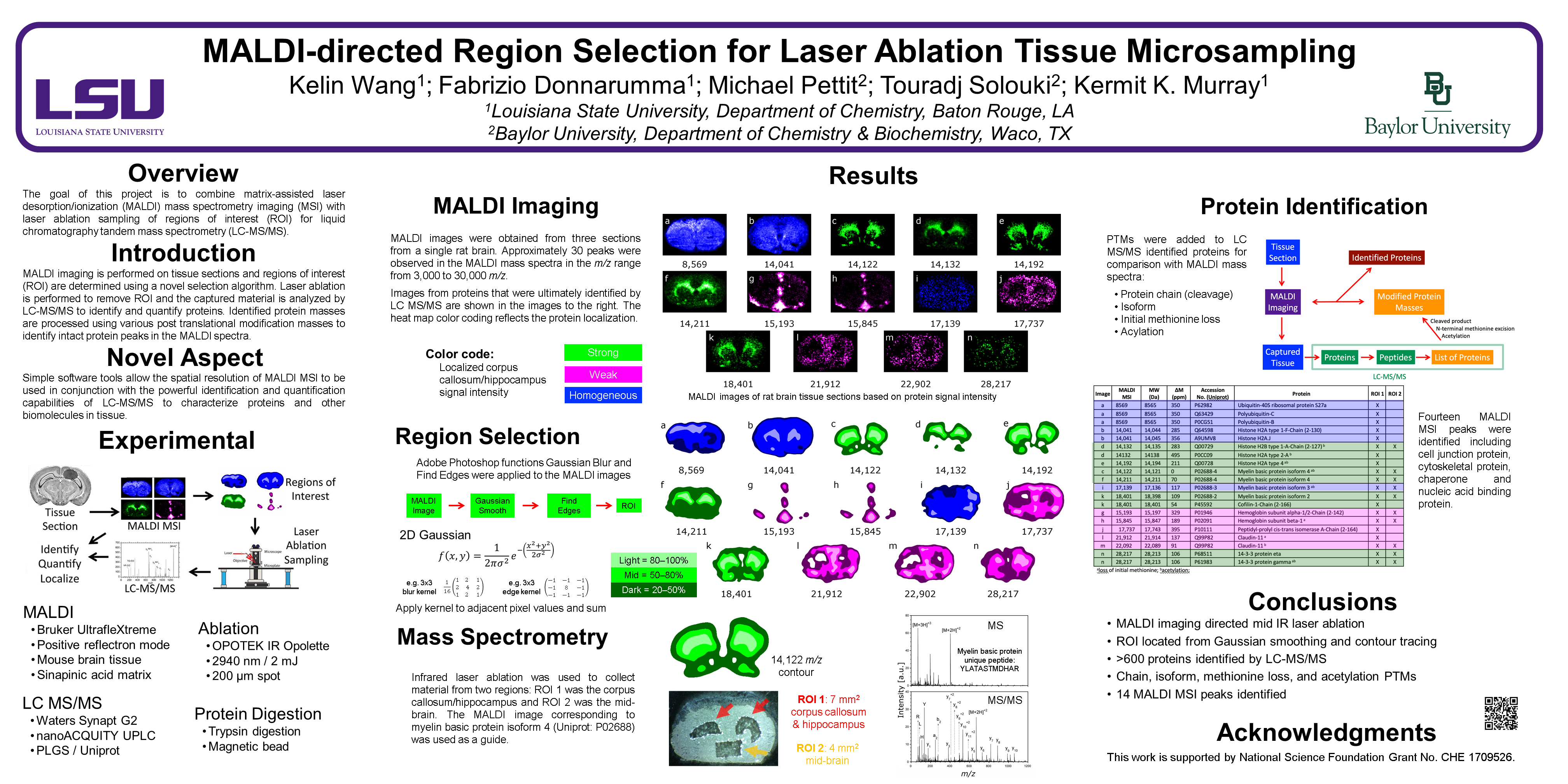
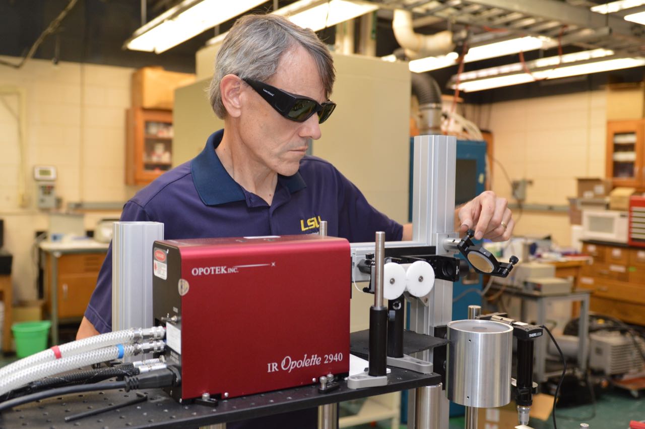
Research group of Kermit Murray at Louisiana State University where we use lasers for sampling and imaging and study the chemistry and physics of laser ablation.
Customer Spotlight – Dr. Kermit Murray – Louisiana State University
R.O. Lawal, F. Donnarumma, K.K. Murray, Electrospray Photochemical Oxidation of Proteins, J. Am Soc. Mass Spectrom. 2019, 30, 11, 2196–2199. /s13361-019-02313-4
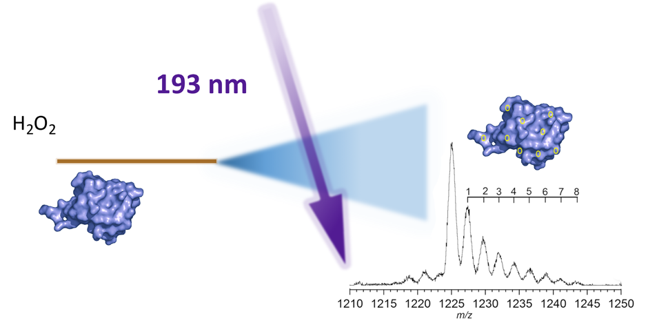
Photooxidation of peptides and proteins by pulsed ultraviolet laser irradiation of an electrospray in the ion source of a mass spectrometer was demonstrated. A 193-nm excimer laser at 1.5-mJ pulse energy was focused with a cylindrical lens at the exit of a nanoelectrospray capillary and ions were sampled into a quadrupole time-of-flight mass spectrometer. A solution containing a peptide or protein and hydrogen peroxide was infused into the spray at a flow rate of 1 μL/min using a syringe pump. The laser creates OH radicals directly in the spray which modify biomolecules within the spray droplet. These results indicate that photochemical oxidation of proteins can be initiated directly within electrospray droplets and detected by mass spectrometry.
B. Banstola, K.K. Murray, “A Nanoparticle Co-matrix for Multiple Charging in MALDI Imaging of Tissue,” Rapid Commun. Mass Spectrom. 16 (2019) 12. doi:10.1002/rcm.8424.
Abstract
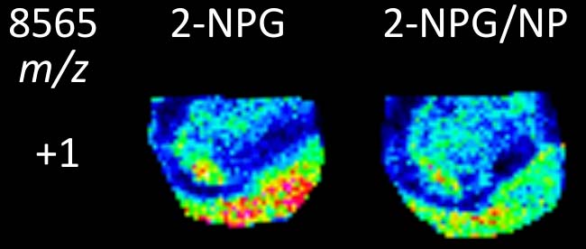
Rationale: A two‐component matrix of 2‐nitrophloroglucinol (2‐NPG) and silica nanoparticles was used for matrix‐assisted laser desorption ionization (MALDI) mass spectrometry imaging of high‐charge‐state biomolecules in tissue. Potential advantages include increased effective mass range and efficiency of fragmentation.
Methods: A mixture of 2‐NPG matrix and silica nanoparticles was applied to cyrosectioned 10 μm thick mouse brain tissue. The mixture was pipetted onto the tissue for profiling and sprayed for tissue imaging. MALDI images were obtained under high vacuum in a commercial time‐of‐flight mass spectrometer.
Results: The combined 2‐NPG and nanoparticle matrix produced highly charged ions from tissue with high‐vacuum MALDI. Nanoparticles of 20, 70, 400, and 1000 nm in diameter were tested, the 20 nm particles producing the highest charge states. Images of mouse brain tissue obtained from highly charged ions show similar spatial localization.
Conclusions: The combined 2‐NPG and nanoparticle matrix produces highly charged ions from tissue through a mechanism that may rely on the high surface area of the particles which can dry the tissue, and their ability to bind analyte molecules thereby assisting in crystal formation and production of multiply charged ions on laser irradiation.

I. Pereira, B. Banstola, K. Wang, F. Donnarumma, B.G. Vaz, K.K. Murray, Matrix-Assisted Laser Desorption Ionization Imaging and Laser Ablation Sampling for Analysis of Fungicide Distribution in Apples, Anal. Chem. 91 (2019) 6051–6056. doi:10.1021/acs.analchem.9b00566.
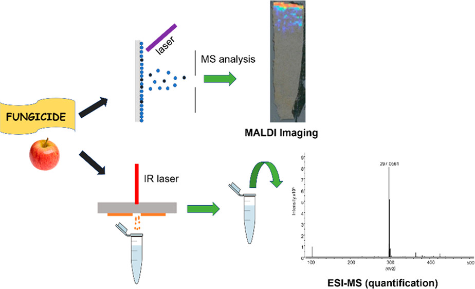
ABSTRACT: A combination of matrix-assisted laser desorption ionization (MALDI) imaging and infrared (IR) laser ablation sampling with offline electrospray ionization mass spectrometry (ESI-MS) was used to determine the distribution of the fungicide imazalil in apples. MALDI images were used to determine the penetration depth of imazalil up to 7 days after its application. IR laser ablation sampling and ESI-MS were used to quantify the rate of penetration of the fungicide, which was determined to be approximately 1 mm per day. Imazalil concentration decreased in the apple skin over the course of the experiment, and after 7 days the fungicide was detected at 0.015 ppm 6 mm inside the apple. Approximately 60% of the pesticide remained in the skin after 7 days. This work demonstrates the utility of MALDI imaging for spatial localization of fungicide in fruit in combination with IR laser ablation and ESI-MS for quantitative analysis.

B. Banstola, C.W. Szot, A.P. Deenamulla Kankanamalage, K.K. Murray, Piezoelectric matrix-assisted ionization, Eur J Mass Spectrom 25 (2019) 202–207. doi:10.1177/1469066718816696
ABSTRACT: We have developed a new actuation method for matrix-assisted ionization with good temporal and spatial resolution using piezoelectric cantilever. A strike from the piezoelectric bimorph cantilever on a thin metal foil was used to remove materials deposited on the opposite side facing the mass spectrometer inlet. Highly charged ions of peptides and proteins were generated from dried droplet deposits and sampled into the inlet of the mass spectrometer. A lateral resolution of 1 mm was obtained with the piezoelectric sampling configuration. Singly charged lipids and gangliosides were detected from tissue with piezoelectric matrix-assisted ionization using a silica nanoparticle co-matrix.

MP 765
Cellular Precision for Infrared Laser Ablation Tissue Microproteomics
Chao Dong; Fabrizio Donnarumma; Kelin Wang; Kermit K. Murray

MOG 03:10pm
Deep-ultraviolet Laser Ablation Sampling for Mass Spectrometry
Remilekun O. Lawal; Fabrizio Donnarumma; Kermit K. Murray

High resolution sampling of biological systems is crucial to revealing the differences in proteins and metabolites amongst heterogeneous cells. Laser ablation sampling for mass spectrometry is a powerful method for analyzing biomolecules in tissue under ambient conditions with high spatial control while eliminating the need for external matrices. It allows off-line analysis by liquid chromatography tandem mass spectrometry and can be combined with mass spectrometry imaging for region of interest selection. The most efficient lasers currently used for ablation sampling use mid-infrared wavelengths that are diffraction limited to spot sizes tens of micrometers in diameter. Short wavelength lasers can be focused to order of magnitude smaller spot sizes for efficient ablation with minimal thermal damage to adjacent sample areas.
WOG 03:30pm
Charge Production by Sublimation of Organic Compounds in Matrix Assisted Ionization
Bijay Banstola; Kermit K. Murray

Matrix assisted ionization (MAI) describes a mode of ionization in which an analyte molecule is mixed with an organic matrix and produces ions when the dried matrix and analyte containing crystals are exposed to external shock or sublime under vacuum. MAI produces highly charged ions from large biopolymers with charge distributions similar to electrospray ionization. When exposed to vacuum, the matrix crystals fracture, which may eject charged particles and clusters containing matrix and analyte or may directly eject highly charged ions. In this study, a method was developed to measure the charge produced during the sublimation of MAI matrix compounds. Sublimation charge production was measured as a function of matrix, crystal size and morphology, pH, and temperature.
WOG 03:30pm
TP 266
TP 362
WP 433
WP 518
Simultaneous Extraction of Proteins, Lipids, and Metabolites for Integrated-omics Approaches for Low Tissue Sampling Volumes
Luke T. Richardson; Amy N. W. Schnelle; Fabrizio Donnaruma; Michael E. Pettit;
ThP 050
ThP 124
Influence of Traumatic Brain Injury on Bile Acid Profiles in the Brains of Rats
Amy N. W. Schnelle; Luke T. Richardson; Fabrizio Donnaruma; Ashok K. Shetty; Kermit K Murray; Touradj Solouki
ThP 413