Kermit K. Murray and Kadeem O. Hayes
- Louisiana State University, Baton Rouge, LA, United States
- Laser Bioanalytics LLC, Baton Rouge, LA, United States
Abstract
A photochemical tissue region modification system for the MALDI imaging workflow.
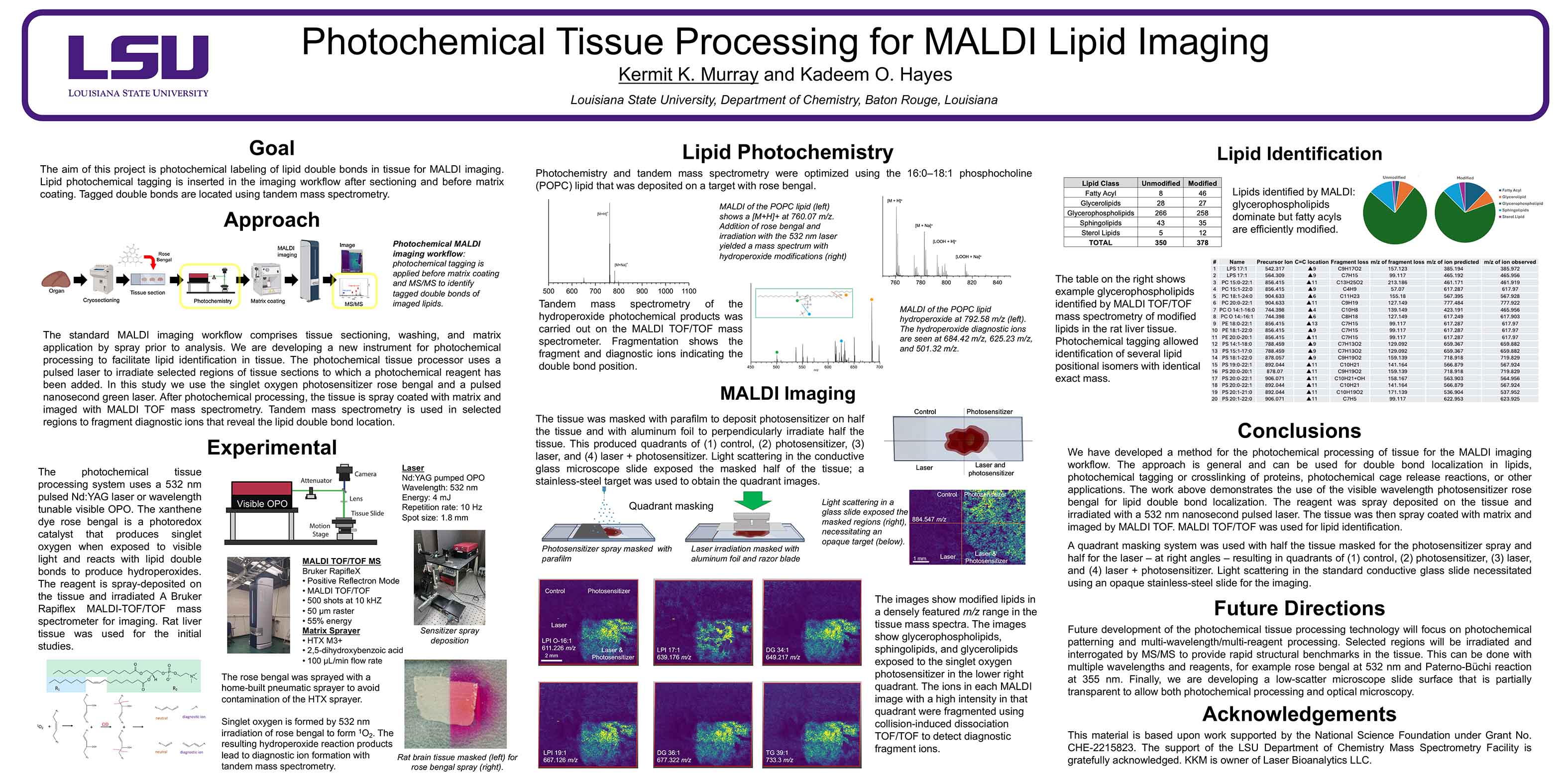
Introduction
The standard MALDI imaging workflow comprises tissue sectioning, washing, and matrix application by spray or sublimation prior to analysis. We are developing a new instrument for tissue photochemical processing that produces biomarker compounds to facilitate compound identification. The current study is aimed at developing a method for lipid double bond localization using MALDI imaging and tandem time-of-flight mass spectrometry. The photochemical tissue processor uses a pulsed laser to irradiate selected regions of tissue sections to which a photochemical reagent has been added. After photochemical processing, the tissue is spray coated with matrix and imaged with MALDI TOF mass spectrometry. Tandem mass spectrometry is used in selected regions to fragment diagnostic ions that reveal the lipid double bond location.
Methods
The photochemical tissue processor is designed to be a standalone device that can be inserted into the MALDI imaging workflow with minimal alteration of the prior and subsequent protocols. The instrument uses a pulsed laser; in the work described below, a 532 nm pulsed Nd:YAG laser or wavelength tunable visible OPO laser were used. The xanthene dye rose bengal is a photoredox catalyst that produces singlet oxygen when exposed to visible light and reacts with lipid double bonds to produce hydroperoxides. The reagent is spray-deposited on the tissue and irradiated, and a Bruker Rapiflex MALDI-TOF/TOF mass spectrometer performs imaging analysis. Rat liver tissue was used for the initial studies.
Preliminary Data
Initial development of the photochemical tissue processor involved optimizing reagent addition, tissue exposure, tandem mass spectrometry, and regionally selective exposure. Optimization of exposure times was performed using the lipid 16:0-18:1 phosphocholine (POPC) which was deposited on a conductive microscope slide target with the rose bengal. It was found that a 10,000 laser shot dosage at 1 mJ per pulse produced >20% lipid conversion. Consecutive 10 µm sections from rat liver were cut using a cryostat microtome at 20 ºC and thaw mounted on conductive microscope slides. Based on the dosage result, the tissue was irradiated for 10 minutes for the imaging studies. A 30 μL volume of 5 mM rose bengal reagent was spray deposited on the tissue using home-built pneumatic sprayer and the carrier solvent allowed to dry. The home-built sprayer avoided contamination of the commercial matrix sprayer, but the workflow will ultimately include a combined reagent and matrix sprayer system. The tissue was irradiated with the laser and vacuum dried after exposure. Next, the 2,5-dihydroxybenzoic acid
matrix was spray deposited. MALDI imaging experiments are performed using a Bruker Rapiflex MALDI-TOF/TOF mass spectrometer. Images were obtained with a raster of 50µm and 500 shots per pixel in positive ion reflection mode with a mass range of 400 – 2000 m/z. Lipids from the rose bengal treated tissue had mass shifts of 32, indicating hydroperoxide formation. The ratio of modified to unmodified lipids increased with time. A total of 94 lipids were identified and one third were modified. Masking of the reagent spray or laser showed that the modified lipids were region localized. Tandem mass spectrometry of diagnostic ions showed headgroup loss and diagnostic ions indicating the double-bond position. Ongoing experiments are aimed at more precise regional lipid modification using laser scanning of specific tissue regions.