Matrix Assisted Laser Desorption Ionization Ion Mobility Time-of-Flight Mass Spectrometry of Bacteria

J.M. Hayes, L.C. Anderson, J.A. Schultz, M.V. Ugarov, T.F. Egan, E.K. Lewis, V. Womack, A. S. Woods, S. N. Jackson, R. H. Hauge, K. Kittrell, S. Ripley, K. K. Murray, “Matrix Assisted Laser Desorption Ionization Ion Mobility Time-of-Flight Mass Spectrometry of Bacteria,” in: C. Fenselau, P. Demirev (Eds.), Rapid Characterization of Microorganisms by Mass Spectrometry, American Chemical Society, Washington, DC, 2011: pp. 143–160. doi:10.1021/bk-2011-1065.ch009.

Abstract
Bacteria IMMS
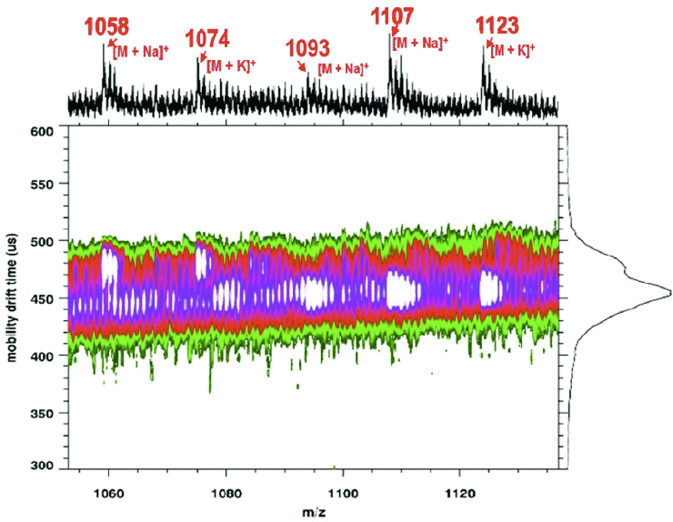
UV MALDI-IM-TOF MS 2-D contour plot of lipopeptide products with Na+ and K+ adducts from whole cell B. subtilis ATCC 6633

Ion mobility mass spectrometry (IMMS) was combined with matrix assisted laser desorption ionization (MALDI) for the analysis of whole cell bacteria. Whole cell Bacillus subtilis ATCC 6633 and Escherichia coli strain W ATCC 9637 bacteria were prepared with a 1:2 analyte to matrix (CHCA) ratio and deposited using the dried-droplet method. Matrix-assisted laser desorption ionization time-of-flight mass spectrometry (MALDI-TOF MS) studies and matrix assisted laser desorption ionization ion mobility time-of-flight mass spectrometry (MALDI-IM-TOF MS) were conducted in parallel to assess the effectiveness of MALDI-IM-TOF MS for microorganism identification. Ribosomal proteins from Escherichia coli strain W ATCC 9637 were observed and assigned using the Rapid Microorganism Identification Database. Isoforms of lipopeptide products and a lantibiotic from the Bacillus subtilis ATCC 6633 species were found in the range 1000–3500 m/z and were identified using MALDI-IM-TOF MS and compared to MALDI-TOF MS data for confirmation. Vacuum ultraviolet (VUV) post-ionization MALDI-IM-TOF MS showed that additional information on lipopeptides could be obtained and used for identification.

Infrared Laser Ablation Sample Transfer for MALDI and Electrospray

S.-G. Park, K.K. Murray, “Infrared laser ablation sample transfer for MALDI and electrospray,” J. Am. Soc. Mass Spectrom. 22 (2011) 1352–1362. doi:10.1007/s13361-011-0163-3.

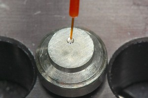
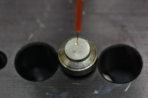
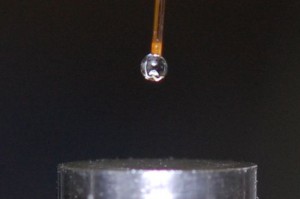
Abstract: We have used an infrared laser to ablate materials under ambient conditions that were captured in solvent droplets. The droplets were either deposited on a MALDI target for off-line analysis by MALDI time-of-flight mass spectrometry or flow-injected into a nanoelectrospray source of an ion trap mass spectrometer. An infrared optical parametric oscillator (OPO) laser system at 2.94 μm wavelength and approximately 1 mJ pulse energy was focused onto samples for ablation at atmospheric pressure. The ablated material was captured in a solvent droplet 1–2 mm in diameter that was suspended from a silica capillary a few millimeters above the sample target. Once the sample was transferred to the droplet by ablation, the droplet was deposited on a MALDI target. A saturated matrix solution was added to the deposited sample, or in some cases, the suspended capture droplet contained the matrix. Peptide and protein standards were used to assess the effects of the number of IR laser ablation shots, sample to droplet distance, capture droplet size, droplet solvent, and laser pulse energy. Droplet collected samples were also injected into a nanoelectrospray source of an ion trap mass spectrometer with a 500 nL injection loop. It is estimated that pmol quantities of material were transferred to the droplet with an efficiency of approximately 1%. The direct analysis of biological fluids for off-line MALDI and electrospray was demonstrated with blood, milk, and egg. The implications of this IR ablation sa

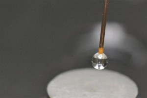
droplet
Solvent droplet suspended from a capillary

A solid-phase bioreactor with continuous sample deposition for matrix-assisted laser desorption/ionization time-of-flight mass spectrometry

J. Lee, S.A. Soper, K.K. Murray, “A solid-phase bioreactor with continuous sample deposition for matrix-assisted laser desorption/ionization time-of-flight mass spectrometry,” Rapid Commun. Mass Spectrom. 25 (2011) 693–699. doi:10.1002/rcm.4921.

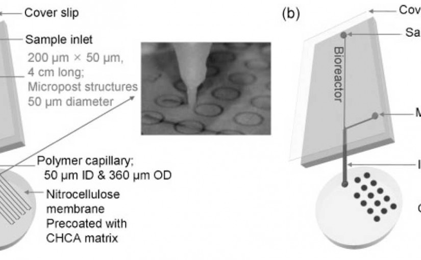
Abstract: We report the development of a solid-phase proteolytic digestion and continuous deposition microfluidic chip platform for low volume fraction collection and off-line matrix-assisted laser desorption/ionization time-of-flight (MALDI-TOF) mass spectrometry. Tryptic peptides were formed in an on-chip bioreactor and continuously deposited onto a MALDI target plate using a motor-driven xyz stage. The bioreactor consisted of a 4 cm × 200 µm × 50 µm microfluidic channel with covalently immobilized trypsin on an array of 50 µm diameter micropost structures with a 50 µm edge-to-edge inter-post spacing. A 50 µm i.d. capillary tube was directly attached to the end of the bioreactor for continuous sample deposition. The MALDI target plate was modified by spin-coating a nitrocellulose solution containing a MALDI matrix on the surface prior to effluent deposition. Protein molecular weight standards were used for evaluating the performance of the digestion and continuous deposition system. Serpentine sample traces 200 µm wide were obtained with a 30 fmol/mm quantity deposition rate and a 3.3 nL/mm volumetric deposition rate.

A schematic of different deposition modes using the PMMA microfluidic chip and immobilized trypsin bioreactor: (a) continuous deposition mode and (b) spot deposition mode. The channel measured 40 mm x 200 mm x 50 mm and had an array of 50 mm diameter posts with a 50 mm inter-post spacing.

Proteome analysis of the leukocytes from the American alligator (Alligator mississippiensis) using mass spectrometry

L.N.F. Darville, M.E. Merchant, A. Hasan, K.K. Murray, “Proteome analysis of the leukocytes from the American alligator (Alligator mississippiensis) using mass spectrometry,” Comp. Biochem. Physiol. Part D Genomics Proteomics. 5 (2010) 308–316. doi:10.1016/j.cbd.2010.09.001.

Abstract: Mass spectrometry was used in conjunction with gel electrophoresis and liquid chromatography, to determine peptide sequences from American alligator (Alligator mississippiensis) leukocytes and to identify similar proteins based on homology. The goal of the study was to generate an initial database of proteins related to the alligator immune system. We have adopted a typical proteomics approach for this study. Proteins from leukocyte extracts were separated using two-dimensional gel electrophoresis and the major bands were excised, digested and analyzed by on-line nano-LC MS/MS to generate peptide sequences. The sequences generated were used to identify proteins and characterize their functions. The protein identity and characterization of the protein function were based on matching two or more peptides to the same protein by searching against the NCBI database using MASCOT and Basic Local Alignment Search Tool (BLAST). For those proteins with only one peptide matching, the phylum of the matched protein was considered. Forty-three proteins were identified that exhibit sequence similarities to proteins from other vertebrates. Proteins related to the cytoskeletal system were the most abundant proteins identified. These proteins are known to regulate cell mobility and phagocytosis. Several other peptides were matched to proteins that potentially have immune-related function.

2D separation of alligator leukocyte stained with ProteoSilver stain for mass spectrometry analysis. Approximately 400 μg of leukocyte protein was loaded and separated on a 2D large gel format.
Primary structure of the 35 kDa lectin protein isolated from American alligator assembled from different endoprotease digestions. The peptide sequences were generated using ESI–MS/MS. Peptides obtained from the different enzymes were highlighted using different colors.

Static Droplet Macro Photos

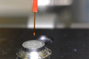
Macro photographs of droplets for laser ablation sample transfer.

Top-view of droplet suspended above laser alblation target

Wide-view of droplet suspended above laser alblation target

Side-view of droplet suspended above laser alblation target, December 2010

Closeup of droplet suspended above laser alblation target

Droplet suspended above laser alblation target

Continuous flow infrared matrix‐assisted laser desorption electrospray ionization mass spectrometry

F. Huang, K.K. Murray, “Continuous flow infrared matrix‐assisted laser desorption electrospray ionization mass spectrometry,” 24 (2010) 2799–2804. doi:10.1002/rcm.4704.

Abstract: Continuous flow infrared matrix-assisted laser desorption electrospray ionization (CF IR MALDESI) mass spectrometry was demonstrated for the on-line analysis of liquid samples. Samples in aqueous solution were flowed through a 50mm i.d. fused-silica capillary at a flow rate of 1–6mL/min. As analyte aqueous solution flowed through the capillary, a liquid sample bead formed at the capillary tip. A pulsed infrared optical parametric oscillator (OPO) laser with wavelength of 2.94mm and a 20Hz repetition rate was focused onto the capillary tip for sample desorption and ablation. The plume of ejected sample was entrained in an electrospray to form ions by MALDESI. The resulting ions were sampled into an ion trap mass spectrometer for analysis. Using CF IR MALDESI, several chemical and biochemical reactions were monitored on-line: the chelation of 1,10-phenanthroline with iron(II), insulin denaturation with 1,4-dithiothreitol, and tryptic digestion of cytochrome c.

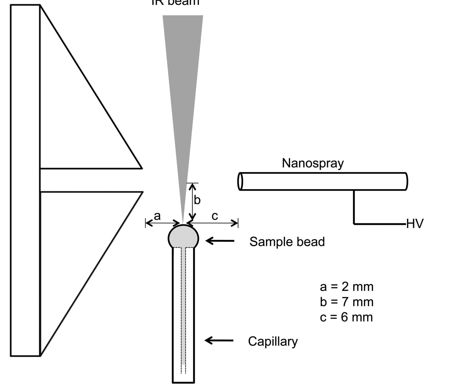
CF IR MALDESI MS interface
Schematic of the CF IR MALDESI MS interface showing the nanospray source, capillary, liquid sample bead, IR laser beam and mass spectrometer orifice (RCM 2010; 24: 2799)
CF IR MALDESI mass spectra of insulin reduction
CF IR MALDESI mass spectra of (a) 500 mM insulin in 50 mM NH4HCO3 water buffer solution, (b) 500 mM insulin mixed with 100 mM 1,4-dithiothreitol (DTT) reducing agent, and (c) denatured insulin b chain after on-line mixing of 500mM insulin with 100mM DTT solution (RCM 2010; 24: 2799)

Glossary of terms for separations coupled to mass spectrometry

K.K. Murray, “Glossary of terms for separations coupled to mass spectrometry,” J. Chromatogr. A. 1217 (2010) 3922–3928. doi:10.1016/j.chroma.2010.03.013.

Abstract: This document is a glossary of terms for separations coupled to mass spectrometry. It covers gas chromatography/mass spectrometry, liquid chromatography/mass spectrometry, and supercritical fluid chromatography/mass spectrometry and the sample introduction, ionization, and data analysis methods used with these combined techniques.

For a complete list of terms, see the Mass Spectrometry Terms Wiki

Mass Spectrometry Terms Wiki

Small molecule ambient mass spectrometry imaging by infrared laser ablation metastable-induced chemical ionization

A.S. Galhena, G.A. Harris, L. Nyadong, K.K. Murray, F.M. Fernandez, Small molecule ambient mass spectrometry imaging by infrared laser ablation metastable-induced chemical ionization, Anal. Chem. 82 (2010) 2178–2181. doi:10.1021/ac902905v.

Abstract

Presented here is a novel ambient ion source termed infrared laser ablation metastable-induced chemical ionization (IR-LAMICI). IR-LAMICI integrates IR laser ablation and direct analysis in real time (DART)-type metastable-induced chemical ionization for open air mass spectrometry (MS) ionization. The ion generation in the IR-LAMICI source is a two step process. First, IR laser pulses impinge the sample surface ablating surface material. Second, a portion of ablated material reacts with the metastable reactive plume facilitating gas-phase chemical ionization of analyte molecules generating protonated or deprotonated species in positive and negative ion modes, respectively. The successful coupling of IR-laser ablation with metastable-induced chemical ionization resulted in an ambient plasma-based spatially resolved small molecule imaging platform for mass spectrometry (MS). The analytical capabilities of IR-LAMICI are explored by imaging pharmaceutical tablets, screening counterfeit drugs, and probing algal tissue surfaces for natural products. The resolution of a chemical image is determined by the crater size produced with each laser pulse but not by the size of the metastable gas jet. The detection limits for an active pharmaceutical ingredient (acetaminophen) using the IR-LAMICI source is calculated to be low picograms. Furthermore, three-dimensional computational fluid dynamic simulations showed improvements in the IR-LAMICI ion source are possible.